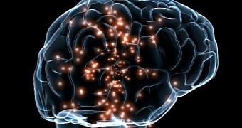
In a recent study in the journal Nature Neuroscience, a team of researchers at the RIKEN Brain Science Institute in Japan describe a novel technique to fashion see-through tissues, entire brains included.
True, such techniques are not exactly breaking news in the world of science. In fact, it was about a year ago that scientists at the California Institute of Technology in Pasadena, US, showed us a transparent mouse that looked more like a deceased jellyfish than a rodent.
Still, the RIKEN Brain Science Institute specialists explain that what makes the technique they developed worthwhile is the fact that, unlike previous ones, it barely causes any damage to biological structures.
Hence, it can be used to research the anatomy of the brain or some other organ without having to slice through it or anything of the kind. “Our method, called ScaleS, is a real and practical way to see through brain and body tissue,” says scientist Atsushi Miyawaki.
Well then, how does it work?
It was a few years back, in 2011, that specialist Atsushi Miyawaki and his research team found that a urea-based aqueous solution could make tissues transparent. The problem with this solution was that it would also eat away at biological structures.
Since then, however, the scientists have found that, by mixing urea with the common sugar alcohol sorbitol in just the right proportion, they can create transparent tissues while maintaining their integrity, Science Daily reports.
They team says they've even tested the technique on animal brains and found that it can make such organs completely see-through while at the same time keeping tissue damage to a minimum.
What's more, they claim their solution can be used to store brains for up to a year without fearing damage. When finally pulled out, the organs are pretty much intact and fit for study, they add.
There are other perks to this technique
Apart from making organs like brains transparent, the solution described by the RIKEN Brain Science Institute specialists can be combined with others to make specific structures glow in certain wavelengths.
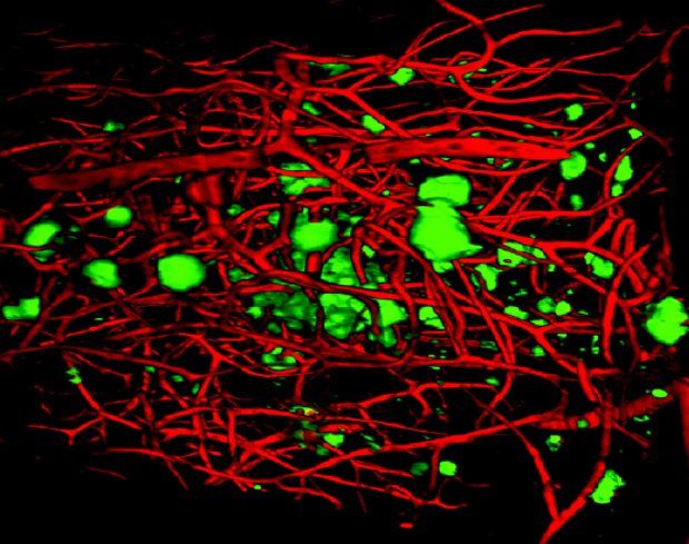
The researchers have even used a few such combinations to study anomalies such as plaques affecting the brains of mice suffering from a variant of Alzheimer's. In the image below, the plaques appear in green and surrounding blood vessels in red.
The experiments proved successful and so the scientists think their technique could very well serve to further our understanding of Alzheimer's and other neurodegenerative diseases of the kind.
“Our technique will be useful not only for visualizing plaques in Alzheimer's disease, but also for examining normal neural circuits and pinpointing structural changes that characterize other brain diseases,” study leader Atsushi Miyawaki said in a statement.

 14 DAY TRIAL //

