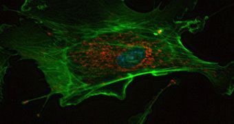
A team of investigators from the University of California in Los Angeles (UCLA) managed to obtain the first-ever 3D images of the entire structure of cells. The impressive achievement was made possible only through the use of an advanced observations instrument called an X-ray diffraction microscope. The new data could be used as a reference point in a wide variety of fields, such as biology and microbiology, bioengineering, nanotechnology and so on. They could also help researchers better understand the mechanisms through which viruses and other pathogens act.
This could, in turn, allow for the creation of novel therapeutic approaches to a wide array of conditions, as well as to the development of new vaccines. The study of biological specimens will also receive an immense boost, given that other observations method are tremendously destructive when used on organic materials. Details of the improvements that led to the recent innovation are published in the May 31 issue of the esteemed journal Proceedings of National Academy of Sciences (PNAS). The researchers also emphasize the differences between the new method and X-ray protein crystallography, which is currently the most commonly-used approach to viewing small molecules.
Unlike proteins, whole cells and other, larger biological structures cannot be easily made into crystals, which means that they generally remain off-limits to scientists. But the new technique, which employs a phenomenon known as lensless imaging, is capable of producing images at resolutions as low as 50 to 60 nanometers. What's even more impressive is the fact that the UCLA group managed to take images of an unstained cell, meaning that the luminescent or fluorescent markers generally used for such studies are no longer needed. Due to this accomplishment, cellular organelles, some viruses and many important protein molecules will from now on become visible as well.
“This is the first time that people have been able to peek into the 3D internal structure of a biological specimen, without cutting it into sections, using X-ray diffraction microscopy. By avoiding use of X-ray lenses, the resolution of X-ray diffraction microscopy is ultimately limited by radiation damage to biological specimens. Using cryogenic technologies, 3D imaging of whole biological cells at a resolution of 5 to 10 nanometers should be achievable. Our work hence paves a way for quantitative 3D imaging of a wide range of biological specimens at nanometer-scale resolutions that are too thick for electron microscopy,” explains UCLA professor of physics and astronomy John Miao.
“Biologists wanted to examine internal structures of the spore, but previous microscopic studies provided information on only the surface features. We are very excited to be able to view the spore in 3D. We can now look into the structure of other spores, such as Anthrax spores and many other fungal spores. It is also important to point out that yeast spores are of similar size to many intracellular organelles in human cells. These can be examined in the future,” adds UCLA assistant researcher in physics and astronomy Huaidong Jiang, the co-lead author of the PNAS paper. Miao was the other coauthor.

 14 DAY TRIAL //