 14 DAY TRIAL //
 Scientists took the first images of the retina in living people and found a surprising diversity among people. Although our perceptions are largely similar, our eyes differ considerably. Thus, scientists argue that the brain is even more involved in vision than previously thought.
One apparently insolvable dilemma about vision is this: How do we know that we are getting the same sensations of color, that my sensation of 'red' is identical to your own sensation of 'red'?
As our editor, Ana Constantinescu writes: "We can imagine a situation where a person has seen, from birth, red instead of green, and green instead of red, but he learned to call red 'green', and green 'red', so even if that person sees the two colors completely reversed, he/she will still act normally. That person will drive through the red and green lights correctly, eat tomatoes and cabbage, and never realize that for him/her cabbage is actually red and tomatoes are green."
However, this dilemma is as not insolvable as it may first seem because colors can be mixed together and various combinations can give the same result. If I would have a sensation of blue and I was taught to call it 'red' and then I mix my 'red' with e.g. yellow (which for the sake of the argument I was educated to call 'yellow'), the end result would be green. Therefore, I would have to be taught to call green 'orange'. However I could obtain the same color by some other combination. The point is that it would be impossible to get a consistent picture of how colors mix together by using some other way of labeling the colors.
The colors are not independent of one another. In fact, there are only three fundamental colors and all the available colors we are experiencing are nothing but combinations of these three. The three fundamental colors are red, blue and green.
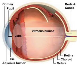
Inside the retina, there are certain cells, called the cones, which are specialized in detecting color. There are only three types of cones: red cones, blue cones, and green cones. Each of these cells absorbs light in a different region of the color spectrum: the red cones absorb red light, blue cones absorb blue light, and green cones absorb green light. Then, they send the information to the brain, and the brain creates the complex visual sensations of colors. It is worth mentioning that we have many sensations of various colors that don't exist in the light spectrum - they are simply brain creations (for example: brown).
This was already known. However, scientists weren't expecting to find such differences between the structures of people's retina. The usual textbook story about the cones in the retina was something like this: "There are 7 million cones in the human retina, 64 percent of which are red, 32 percent green, and 2 percent blue." This was found to be false.
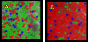
The new study showed there is enormous variability, sometimes up to 40 times, in the relative number of green and red cones in the retina. In this image there are two pictures of the retina taken from two different persons. The person on the left has more green cones than red, while the person on the right has much more red cones than green cones. Nevertheless, they perceive colors likewise. None of them has any vision abnormality.
"[This] suggests that there is a compensatory mechanism in our brain that negates individual differences in the relative numbers of red and green cones that we observed," said Joseph Carroll, a researcher at Center for Visual Science at University of Rochester and a collaborator of the study.
In order to obtain the pictures of the retina the researchers used a technique called 'adaptive optics imaging' first developed by astronomers.
"Adaptive optics is a technique borrowed from astronomy where it is used to obtain sharp images of stars from telescopes on the ground," said David Williams, Director of Center for Visual Science at the University of Rochester. "All such telescopes suffer from blur due to the effects of turbulence in the Earth's atmosphere. In our case, optical defects in the cornea and lens of the eye blur images of the retina."
Williams and colleagues corrected the 'defects in the cornea' by using deformable mirrors, which bend and morph according to each person's eye. This allowed them to see and map single cells such as the cones. The researchers hope to use the same techniques to better understand various forms of color blindness and different kinds of retinal disease.
Photo credit: University of Rochester