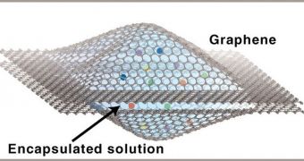
Physicists in the United States have taken an important step forward toward enabling the scientific community to observe physical, chemical and biological phenomena at the nanoscale in real-time. Recently, they have produced a series of videos showing how platinum nanocrystals grow.
The work was carried out by experts at the US Department of Energy's (DOE) Lawrence Berkeley National Laboratory (Berkeley Lab) and the University of California in Berkeley (UCB).
Their achievement breaks a number of firsts, since the video clips depict the growth of platinum nanocrystals between two sheets of graphene, in real time and at the atomic scale. This could lead to a much better understanding of previously mysterious phenomena.
Basically, what the researchers did was create a method of encapsulating liquids containing nanoscale crystals between layers of the 2D carbon compound graphene, the world's toughest material. This enabled the scientists to study the nanocrystals using an electron microscope.
“Watching real-time chemical reactions in liquids at the atomic-scale is a dream for chemists and physicists,” explains team member Jungwon Park. He is based at the Berkeley Lab Materials Sciences Division (MSD) and the UCB Chemistry Department.
“Using our new graphene liquid cell, we’re able to capture a small amount of liquid sample under a high vacuum condition for taking real-time movies of nanoparticle growth reactions,” he adds.
“Since graphene is chemically inert and extremely thin, our liquid cell provides realistic sample conditions for achieving high resolution and contrast,” Park says. He was the lead author of a paper entitled “High-Resolution EM of Colloidal Nanocrystal Growth Using Graphene Liquid Cells.”
The work appears in the April 20 issue of the top journal Science. UCB researcher Jong Min Yuk was also a co-lead author of the paper. Experts from the Center of Integrated Nanomechanical Systems and the Advanced Institute of Science and Technology (KAIST) in Korea were also a part of the team.
An electron microscope was used for this study because this type of instrument can see objects and structures several thousand times smaller than an optical microscope can. However, the former can only operate in vacuum conditions.
The DOE Office of Science and the National Research Foundation of Korea provided the necessary funds for this investigation.

 14 DAY TRIAL //