 14 DAY TRIAL //
 There have been many debates and studies on the HIV virus and the AIDS disease, which wreak havoc around the globe. Researchers are starting to make progress toward developing a vaccine and other advanced prevention methods, but their efforts are not always successful. This should come as no surprise considering that up until now, they have been unable to see how the virus looks in three dimensions.
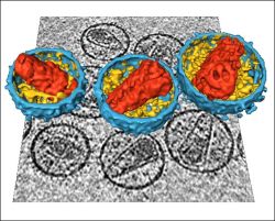
It took the effort of a team of Oxford, Heidelberg and Munich researchers to obtain the first 3D image of the virus which strips the immune system of all its power.
Normal microscopes are unable to distinguish an organism 60 times smaller than a red blood cell, and the electron ones or X-rays are able to "see" it, but the results are often unsatisfactory because the virus varies so much in size and shape.
Professor Stephen Fuller from Oxford and his colleagues used a technique called cryo-electron tomography to look in detail at the morphology of the virus. The technique has been used to see the virus before, but only for 2D images. The researchers took images from hundreds of different angles and combined them with the computer.
An HIV particle, like any virus, is not a cell but rather is strands of genetic code wrapped in protein. Viruses invade living cells and take them over by usurping the cell's genetic code with the virus's genetic code.
It seems that the HIV virus's ability to change its size and shape makes it so efficient. One of the researchers' biggest dilemmas is the way in which it manages to change the shape without destroying its structure.
The 3D image seems to explain this mystery. Instead of the central region of the virus organizing its growth, as in most viruses, the virus membrane and the core interact so that the core stops growing only when it reaches the membrane's limit. The inner surface of the viral membrane 'directs' growth, which keeps the important parts of the structure consistent whilst allowing size variation.
"Identifying how the virus grows will allow us to address the formation of this important pathogen and understand how it accommodates its variability. This could inform the development of more effective therapeutic approaches," Professor Stephen Fuller said.