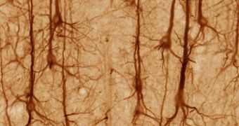
In an achievement that could help researchers analyze the complexity and variety of nerve cell connections developing in the human brain, experts in the United States managed to develop a method for locating and counting synapses that are created between neurons.
Each of the nerve cells can connect to thousands of other neurons, producing network that are extremely complex, and which underly most of our mental abilities. With the help of a state-of-the-art imaging system, scientists were recently able to accurately locate and count these connections.
The team, which is based at the Stanford University School of Medicine, used brain tissue samples collected from mice to conduct the investigation. In addition to observing the number of synapses present in the samples, the method also helped experts catalog their variety.
According to previous studies, the average human brain contains in excess of 200 billion neurons, which is about twice as much as how many stars are in the Milky Way. These cells are interconnected via hundreds of trillions of synapses.
The basic role of these contact points is to act as relays. Electrical signals passing through one neuron eventually make their way to synapses, where they are relayed to the next neuron. This is how impulses from the cortex make their way to the rest of the body.
According to Stephen Smith, PhD, each nerve cell can create as many as tens of thousands of connections with the neurons around it, which means that synapses are packed very closely together, and exist on numerous levels.
As such, keeping track of their complexity and number has been a pain thus far. However, understanding that is key to making sense of the complex neuronal circuits and pathways that allow us to move, feel and think.
With the new approach, scientists may soon become able to map this complex network, says Smith, who is a professor of molecular and cellular physiology at Stanford. He is also the senior author of a research paper on the issue, which appears in the November 18 issue of the esteemed journal Neuron.
He explains that the new imaging method is tremendously complex itself. Its two main components are high-resolution photography and specialized fluorescent molecules. The latter bind to various proteins and enzymes, and then glow in different colors.
As this happens, a powerful computer picks up the data, and then converts the information into imagery, allowing researchers to peer deep into synaptic networks. They can then make sense of how neural connections are organized.
“In a human, there are more than 125 trillion synapses just in the cerebral cortex alone. Now we can actually count them and, in the bargain, catalog each of them according to its type,” Smith explains.
“I anticipate that within a few years, array tomography will have become an important mainline clinical pathology technique, and a drug-research tool,” the expert concludes.

 14 DAY TRIAL //