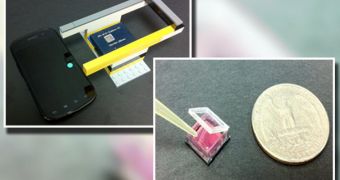
The standard Petri dish has been in use among scientists since the late 19th century, helping them monitor how bacteria grow, and also if people are infected with a certain pathogen or not. Now, a team of scientists in the United States create an advanced version of the tool, and named it ePetri.
The group, based at the California Institute of Technology (Caltech), in Pasadena, says that the new device does away with the need to extract the petri dishes out of the incubators every time a test needs to be performed. Until now, this is how things were done.
Cultures placed in these dishes are generally grown in an incubator, so as to accelerate the development of bacteria. At various intervals, the instruments are extracted and then placed under a microscope, where a scientist assesses the evolution of bacterial cultures.
This approach to using the petri dish is very complex and time consuming, the Caltech team says. Experts would be better off spending their time doing something else than babysitting bacterial cultures. This is where ePetri comes in.
According to its creators, the main thing that separates the new instrument from standard dishes is the fact that it can image cell growth continuously and autonomously, while delivering whatever data it collects directly to a computer.
“Our ePetri dish is a compact, small, lens-free microscopy imaging platform. We can directly track the cell culture or bacteria culture within the incubator,” Caltech electrical engineering graduate student and lead study author Guoan Zheng explains.
“The data from the ePetri dish automatically transfers to a computer outside the incubator by a cable connection. Therefore, this technology can significantly streamline and improve cell culture experiments by cutting down on human labor and contamination risks,” the expert says.
Details of how ePetri works were published in this week's online issue of the esteemed journal Proceedings of the National Academy of Sciences (PNAS). In addition to reducing human labor time, the new dishes also eliminate the need to have large microscopes available at all times.
Interestingly enough, the device itself is built out of a Google smartphone, an image sensor from a commercially-available cell phone, and some LEGO parts. Wires running from the image-sensor chip are used to take data from inside of the incubator to a computer outside.
“Until now, imaging of confluent cell cultures has been a highly labor-intensive process in which the traditional microscope has to serve as an expensive and suboptimal workhorse,” Changhuei Yang adds.
“What this technology allows us to do is create a system in which you can do wide field-of-view microscopy imaging of confluent cell samples. It capitalizes on the use of readily available image-sensor technology, which is found in all cell-phone cameras,” the expert argues.
Changhuei Yang was the senior author of the PNAS paper. He holds an appointment as a professor of electrical engineering and bioengineering at Caltech.

 14 DAY TRIAL //