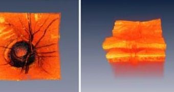
The retina is one of the most fragile tissues of our organisms.
So fragile, that the projects of the bionic eyes try to eschew it. But this is the tissue where images are captured and sent as electrical signals to the brain and in fact, the cause of almost all types of blindness.
In order to improve the diagnoses of many ocular diseases, MIT researchers have worked on a new type of laser for taking 3-D high-resolution images of the retina.
The new technique works on Optical Coherence Tomography (OCT), which employs light to get high-resolution, cross-sectional images of the eye to detect extremely subtle cell changes in retinal disease.
OCT was first developed in the early 1990s by a team led by MIT Professor James Fujimoto; he is also the author of the new research. "Within the last few years optical coherence tomography has become a standard diagnostic for ophthalmology. New techniques are now enabling dramatic increases in image acquisition speeds. These advances promise to enable new and powerful three-dimensional visualization methods which could improve early diagnosis of disease and treatment monitoring," said Fujimoto, from MIT's Department of Electrical Engineering and Computer Science and the Research Laboratory of Electronics.
Conventional OCT technologies use series of 2-D (two dimensional) cross-sectional retinal pictures, which can be employed through combinations to get 3-D images.
The OCT systems scan light back and forth across the eye, and the echo time delay of reflected light allows the building of high-resolution images.
Current OCT systems can scan the eye at speeds of several hundred to several thousand lines per second, while humans can keep the ocular globe still for just about one second, thus there is a low level of three-dimensional data achieved by commercial OCTs. The new laser effectuates retinal scans at rhythms of up to 236,000 lines per second, 10 quicker than conventional technology.
Further development could enable physicians to get three-dimensional "OCT snapshots" of the eye, a crucial tool for earlier and improved diagnoses of retinal diseases like diabetic retinopathy, glaucoma and macular degeneration.

 14 DAY TRIAL //