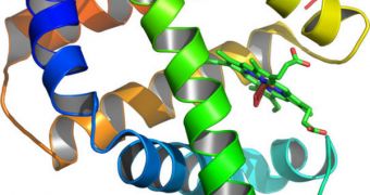
Proteins are arguably the most important molecules in the human body, as they control everything a cell does. Therefore, studying them is critically important, but equally as difficult. An innovation in microscopy is now making it easier for scientists to investigate these elements.
Viruses, genes and neurons all have something in common, and that is the fact that the way they function is regulated by proteins. But many of these molecules – that have different functions – look exactly the same chemically.
However, they are not the same. Two proteins may share the same chemical composition, but their functions may be different on account of their external appearance. The angles at which they are bent are equally important at determining their role as the chemicals they contain are.
Experts with the Rockefeller University recently developed a new way of using polarized light to make protein studies a lot easier. Their method enables the study of how a certain molecule is oriented inside a living cell.
This line of work is tremendously important because it covers a gap in knowledge that was left behind by other, less in-depth imaging technologies. For their new study, the researchers focused on a large cluster of proteins called the nuclear pore complex.
The NPC's main role is to regulate which chemicals can pass through to cell's nucleus. It basically sets some conditions that molecules seeking to enter must fulfill before they are allowed passage.
“Our new technique allows us to measure how components of large protein complexes are arranged in relation to one another,” explains the head of the Rockefeller Laboratory of Cellular Biophysics, expert Sandy Simon.
“This has the potential to give us important new information about how the nuclear pore complex functions, but we believe it can also be applied to other multi-protein complexes such as those involved in DNA transcription, protein synthesis or viral replication,” she goes on to say.
Polarized light microscopy conducts studies in exact the resolution area that is omitted by both electron microscopy and X-ray protein crystallography. While the former can image the broader context of a protein, the latter can see only minute details.
Using the new approach, experts will be able to observe the protein, its orientation and its structure in more detail than ever before.
“Our experimental approach to the structure is synergistic with other studies being conducted at Rockefeller, including analysis with X-ray crystallography in Günter's lab and electron microscopy and computer analysis in Mike Rout's lab,” Simon adds.
“By utilizing multiple techniques, we are able to get a more precise picture of these complexes than has ever been possible before,” she concludes.

 14 DAY TRIAL //