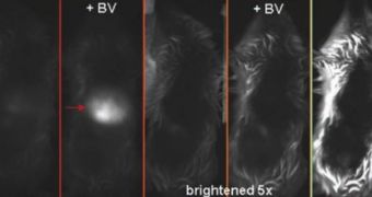
The thing that sets this fluorescent protein apart from all others that are currently used in similar researches is the fact that the extremophile kind glows not in the visible light spectrum, but in infrared. This means that it also remains visible when deeply immersed inside the cell it's set to reveal to researchers. While visible light cannot go through obstacles such as cellular walls, infrared wavelengths can, and, therefore, a very wide array of processes that have remained hidden to bioengineers until now could soon be revealed. Basically, researchers say that the new molecule will allow them to tag specific proteins in living mammal cells, and thus gain insight into some of their organisms' most hidden secret processes.
“Because their wavelengths penetrate tissue well, infrared-fluorescent proteins are suitable for whole-body imaging,” Biochemists Roger Tsien and Xiaokun Shu, from the University of California in San Diego (UCSD), explained in the Thursday issue of the journal Science. Tsien is famous worldwide for his work with the green fluorescent protein, also known as GFP, which was first used to tag only certain types of genes in the human body. Over the years, the expert's lab has developed thousands of derived markers, which can now be attached to any gene in the body.
What's even more amazing about the new infrared protein is the fact that the team don't use it in the basic forms in which they harvest it from the Deinococcus radiodurans extremophile microbe. That is to say, in it's natural state, the molecule is simply too dim to be of any use in scientific research, so Tsien and Shu have augmented it with a few amino-acids, changing its regular composition. This has resulted in a much brighter protein, able to trace the intricate developments going on inside living animal cells.
Scientific investigations are also made easy by the use of a fluorescence molecular tomograph (FMT), which is a scientific observation device that is able to render 3D images from 2D scans. The way it does that is by taking scans of a certain target at various depths from the same angle, and then combining them in a single image. This results in a multi-layered image, which can then be joined by others, to create a complete view of a liver, a kidney, a lung, or any other organ in the human body.
Shu and Tsien have also announced that an additional 1,500 molecules similar to the one they've discovered have already been identified, and that refining FMT to the level of GFP scans could result in increased advancements in bioengineering. “The use of fluorescent proteins in intact animals, such as mice, has been handicapped,” they told about the progress recorded so far, Wired reports.

 14 DAY TRIAL //