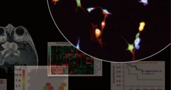
Over the past few years, science has made numerous advancements in figuring out some of the most essential pathways associated with the development of cancer. This condition affects millions of people every year, and investigators are desperate to understand it better. Thus far, scientists have identified a large number of biomarkers, substances which can indicate the presence of cancer in a targeted cell or tissue. But now, a new device promises to innovate this field of investigations.
Researchers at the University of California in Los Angeles (UCLA) have recently developed a novel microfluidic image cytometry (MIC) platform, a device that combines all the functionality of a microfluidic device and microscopy-based cell imaging into a single apparatus. The UCLA team says that its machine can accurately measure cell-signaling pathways that occur in cancer tumors at a single cell level. The innovative platform is capable of producing its results after analyzing only small samples of clinical tissue.
“The MIC is essentially a cancer diagnostic chip that can generate single-cell 'molecular fingerprints' for a small quantity of pathology samples, including brain tumor tissues. We are exploring the use of the MIC for generating informative molecular fingerprints from rare populations of oncology samples – for example, tumor stem cells,” explains UCLA associate professor of molecular and medical pharmacology Dr. Hsian-Rong Tseng. The expert was also one of the leaders in the new investigation.
The fingerprints Tseng was referring to are actually molecular measurements of target samples, collected in vitro. “Because the measurements are at the single-cell level, computational algorithms are then used to organize and find patterns in the thousands of measurements. These patterns relate to the growth signaling pathways active in the tumor that should be targeted in genetically informed or personalized anticancer therapies,” explains UCLA assistant professor of molecular and medical pharmacology, Thomas Graeber. Tseng and Graeber, both of whom are also based at the California NanoSystems Institute (CNSI), were co-leaders on the investigation.

 14 DAY TRIAL //