A group of experts in the United States announces the development of a new method for detecting liver cancer, which they say could help oncologists discover the disease earlier on than currently possible.
The approach can detect cancer tumors that are as small as 5 millimeters, a significant improvement from other available techniques. The work that led to this innovation was carried out by experts at the Brown University.
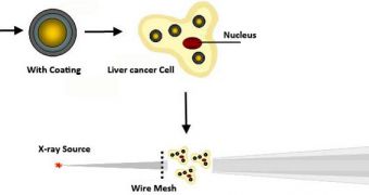
Experts here say that the most important components of their approach are gold nanoparticles, which they use to improve the resolution of X-ray imaging technologies. These agents can help scatter the X-ray in more relevant patterns, uncovering smaller tumors than other imaging techniques can.
This work is very important because more than half a million people are diagnosed with hepatocellular carcinoma – the most common type of liver cancer – every year. Most of these individuals are located in Southeast Asia and sub-Saharan Africa.
The prognosis is very bleak for these people. The disease kills swiftly, and most of those who develop it die within 6 months of diagnosis or less. As such, detecting the cancer earlier on becomes an extremely important objective.
In a new paper, appearing in the esteemed journal Nano Letters, a publication of the American Chemical Society (ACS), the team explains that other detection methods tend to discover liver cancer only when the tumors have already grown beyond 5 centimeters.
At this point, the disease is extremely aggressive, resilient to the effects of chemotherapy and radiotherapy, and extremely difficult to remove via surgery. These factors contribute to the grim outcome most liver cancer patients have.
But the Brown team says that its approach could help improve the outlook. Experts here coat gold nanoparticles with a charged polymer, and then insert them into the liver. An X-ray scatter imaging technique is tend used to image the tissue.
In lab experiments, experts were able to discover tumors as small as 5 millimeters. “What we’re doing is not a screening method,” explains Brown University professor of chemistry and corresponding paper author Christoph Rose-Petruck.
“But in a routine exam, with people who have risk factors, such as certain types of hepatitis, we can use this technique to see a tumor that is just a few millimeters in diameter, which, in terms of size, is a factor of 10 smaller,” he adds.
The new research was sponsored by the Brown Institute for Molecular and Nanoscale Innovation, the US National Institutes of Health and the Department of Energy (DOE).

 14 DAY TRIAL //