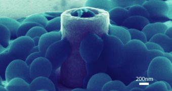
Researchers at the US Department of Energy's (DOE) Lawrence Berkeley National Laboratory (Berkeley Lab), in California, announced recently that they were working on a new way of preventing bacterial infections in a wide array of settings. Their approach does not rely on using antibiotics.
The work was prompted by the ever-growing incidence of cases where bacteria such as Staphylococcus aureus (S. aureus) and methicillin-resistant S. aureus (MRSA) infect medical equipment in hospitals, as well as new prosthetic joints or artificial heart valves in transplant patients. Staph infections are always dangerous and can even become life-threatening.
As the bug evolves resistance to antibiotics, drug designers are working on new chemicals to counteract MRSA. However, the Berkeley Lab is taking a different approach, one that would fight the bacteria by simply not allowing the infection to begin in the first place. In order for microorganisms including S. aureus to thrive, they first need to attach themselves to materials or organs.
This is where the new study comes in. Berkeley Lab investigators are currently working on creating metallic nanostructures capable of preventing MRSA cells from attaching to their surface. The first step in their research was to figure out how S. aureus affixes itself to the material.
For this purpose, the team used structures of varied shapes and sizes, and looked at how the bacterial cells approached their colonization. This line of study has significant implications for medical care.
“By understanding the preferences of bacteria during adhesion, medical implant devices can be fabricated to contain surface features immune to bacteria adhesion, without the requirement of any chemical modifications,” researcher Mohammad Mofrad explains.
He holds an appointment as a faculty scientist with the Physical Biosciences Division at the Berkeley Lab and is also a professor of bioengineering and mechanical engineering at the University of California in Berkeley (UCB). Details of the research he conducted appear in a recent online issue of the esteemed journal Biomaterials.
After creating nanomaterials of different types, the team used an imaging technique called scanning electron microscopy (SEM) to investigate which shape better rejected the S. aureus cells. When the material resembled tubular-shaped pillars with holes in them, the bacteria survived best. The worst-case scenario for the superbug was a cylindrical pillar without any holes in it
“The bacteria seem to sense the nanotopography of the surface and form stronger adhesions on specific nanostructures,” explains the lead author of the research, PBD expert Zeinab Jahed. He is also a graduate student in the UCB Molecular Cell Biomechanics Laboratory, under Mofrad. Scientists at the University of Waterloo, in Canada, were also a part of the research effort.

 14 DAY TRIAL //