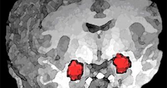
Stanford University investigators discovered the neural pathways that are responsible for controlling anxiety in the brains of unsuspecting mice, and are convinced that they could translate the findings to humans as well.
Brain cell connections determining anxiety-related behavior are tremendously intricate, the team says, and it was only through sheer determination and laborious research that experts made their discoveries.
In the experiments, experts used genetically-engineered lab animals, whose neurons could be activated using light. They then shone a light source on these nerve cells, and observed precisely which of them is involved in activating or deactivating anxiety-related behaviors.
Such studies are tremendously important for humans, because anxiety disorders represent the most common class of psychiatric disease, and they affect millions of people around the world.
Developing new treatments, therapies, cures or drugs to address this array of disease is very difficult without pinpointing the exact origin that anxiety has in the human brain, says Karl Deisseroth.
He is the leader of the Stanford research team, and holds an appointment as an associate professor of psychiatry and behavioral sciences and bioengineering at the university.
The expert and his team were able to identify two critically-important neural pathways in the brain. The first was found to be involved in promoting anxiety, while the second one was proved to reduce it.
Both of them are found in the amygdala, a region of the brain that has been demonstrated to play an important part in controlling fear, the body's fight or flight response, and other primary feelings.
In a paper detailing the study – published in the latest issue of the top scientific journal Nature – the experts explain how they used a research technique called optogenetics to find the two pathways.
What this approach allows experts to do is manipulate the way in which single neurons in the brain of living animals fires, or otherwise conducts its regular activity. Additional engineering was used to make the nerve cells produce a light-controlled protein.
When certain wavelengths of light are shone on them, these proteins get activated, as do the nerve cells containing them. This enables scientists to fine-tune neural activity in any pathway they choose.
“Fear and anxiety are different. Fear is a response to an immediate threat, but anxiety is a heightened state of apprehension with no immediate threat,” Deisseroth explains.
“They share the same outputs, for example physical manifestations such as increased heart rate, but their controls are very different,” he goes on to say.
“Now that we know that these cell projections [in the amygdala] exist, we can first use this knowledge to understand anxiety more than we do now,” the lead investigator adds.
This study was made possible by funding from the National Alliance for Research on Schizophrenia and Depression, the US National Institutes of Health (NIH) and the National Science Foundation (NSF).

 14 DAY TRIAL //