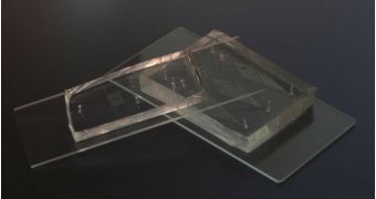
Experts with the Georgia Tech School of Chemical & Biomolecular Engineering announce the development of a new type of microfluidic device, that could make studying various embryos in more exquisite detail than ever before.
The instrument is capable of orienting hundreds of fruit flies embryos, as well as other embryos, in a manner that allows scientists to study the evolution of individuals with various genetic backgrounds at the same points in time.
When the embryo develops in the flies, the most important thing that forms is the dorsoventral axis, a filament-like structure that runs from the back to the bell. How various molecules work together to form this structure is still unknown.
Expert do know that certain proteins are involved in the process, but they have yet to determine the precise interplay that allows for the axis to develop the same in all individuals.
This is why it's so important to analyze offspring from large number of parental pairs. The varied genetic background causes particularities for each individual, but the common mechanism of dorsoventral axis development should be common among all.
If that is true, then the said mechanism should make itself obvious if numerous embryos are analyzed at various points in time during their development. The new microfluidic device allows the researchers to perform this operation with ease.
“Collecting and analyzing the signaling and transcriptional patterns of the dorsoventral axis typically requires manual manipulation of individual embryos to stand them on their ends, making it difficult to conduct high-throughput experiments that can achieve statistically significant results,” says Hang Lu.
The expert is an associate professor in the Georgia Tech School of Chemical & Biomolecular Engineering, Science Blog reports. The device he helped create can handle several hundred fruti fly embryos in a matter of minutes.
“Experimentally, we found on average 90 percent of the embryos became trapped in the device, which will be valuable for studies that only have a small number of embryos available,” Lu explains.
Each of the new microfluidic devices contains about 700 embryo traps, which are located alongside an microscale, S-shaped central channel.
“The trap array device provided a significant increase in the number of fixed and live embryos we could image simultaneously and allowed us to accurately resolve issues of interest to developmental biologists today,” Lu adds.
Details of the device were published in the December 26 advance online issue of the esteemed scientific journal Nature Methods. The National Science Foundation (NSF) and the National Institutes of Health (NIH) supported the research.

 14 DAY TRIAL //