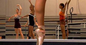
Researchers at the University of California in Irvine announce the development of a new Magnetic Resonance Imaging (MRI) technology, that allows for the observations of ballet dancers en pointe.
It takes a lot of skill, effort, training and patience to reach the degree of physical performances that allows for en pointe dancing, which is when ballerinas only touch the ground with the tip of their feet.
The position is extremely demanding for them, which is why the injury rate among professional ballet dancers is so high.
With the aid of the new MRI method, UCI athletic trainer Jeff Russel hopes to be able to develop new treatments against injuries. Additionally, the data could also be used to develop better prevention methods.
“The physical rigors of ballet are as great as in any other sport, and participants have the injury rate to prove it,” the expert says. He is now an assistant professor of dance science at UCI.
“Dancers endure long rehearsal periods and repetitive body movements. They also tend to have a higher pain threshold and tolerance for pain than nondancers. Consequently, they experience a lot of injuries,” the scientist explains further.
In addition to tendon and muscle strains, snapping hips, damaged Achilles' heels, and stress fractures are among the most common form of damage incurred by ballet dancing.
In the latest issue of the esteemed medical journal Acta Radiologica, the expert and his group presented the new MRI method, which can be used to evaluate parameters belonging to the health of the foot muscles and tendons as the dancers sit en pointe.
But the task is very demanding for the dancers themselves, given that they have to maintain this tremendously complex posture for about two and a half minutes.
The advantage is that their weight-bearing anatomy is exposed, allowing researchers like Russel to determine the points that are most at risk of experiencing issues related to dancing.
The research team demonstrated that using MRI is possible in the most strenuous conditions. Russel showed that, if the situation demands it, people can use wooden blocks to align the dancers' feet with the scanner.
An open MRI table is used for the job. It is placed vertically, and made to rotate around the dancer. This allows experts to produce an image of how muscles and bones reposition themselves within the ankles.

 14 DAY TRIAL //