Rice University investigators announce the creation of a new diagnostics method for Alzheimer's disease, which functions in a very straightforward manner. The team developed a light-switching complex that can attach itself to the amyloid protein, a remarkable scientific feat.
The amyloid protein tends to clump up in people who develop neurodegenerative forms of dementia such as Alzheimer's. By tracking the new complex, experts can keep track on how this specific protein behaves in the brain of patients.
When these molecules begin to clump up together, the intensity of the feedback that the new complex produce will increase, alerting doctors to the presence of abnormal amyloid accumulations in the brain.
In fact, the research team believes that the early detection of this dangerous disease might become extremely simple in the future. Rice bioengineer Angel Martí led the research team that developed the new diagnostics approach.
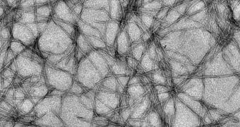
He and his group directed their attention on a specific class of metallic molecules, which are known for their propensity to seek out and attach themselves to fibrils. These are structures in the brain that are formed by amyloid protein plaques; they are a hallmark of Alzheimer's.
The metallic molecules are basically complexes of dipyridophenazine ruthenium, whose luminescence can increase 50 times over when they attach themselves to amyloid fibrils. As such, they act as a lighthouse within the brain, indicating when the fibrils are.
By analyzing this feedback, neurologists will soon be able to tell whether a patient only has the normal amount of amyloid proteins in their brain, or if plaques are beginning to form. By participating in regular controls, people will allow doctors to discover the disease as soon as the first symptoms appear.
“The researchers said their complexes could be fitting partners in a new technique called fluorescence lifetime imaging microscopy, which discriminates microenvironments based on the length of a particle's fluorescence rather than its wavelength,” a Rice press release reads.
By applying a technique called “time gating” to the spectroscopic assays obtained by shining light on the new molecules, experts can weed out wavelengths that are only produced between specially-selected time intervals.
“We specifically took the values only from 300 to 700 nanoseconds after excitation. At that point, all of the fluorescent media have pretty much disappeared, except for ours,” says Rice graduate student Nathan Cook, the lead author of the new research.
“The exciting part of this experiment is that traditional probes primarily measure fluorescence in two dimensions: intensity and wavelength. We have demonstrated that we can add a third dimension – time – to enhance the resolution of a fluorescent assay,” he concludes.

 14 DAY TRIAL //