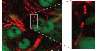
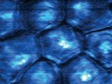
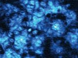
Detecting extremely small molecules that have no fluorescence is very difficult to accomplish, especially if you don't know what you're looking for. Scientists at the Harvard University have recently managed to break this limitation, when a team led by expert X. Sunney Xie has created a new microscopic technique that allows for the identification of previously unseen molecules. The method also works for imaging in color and in great detail molecules that have no natural fluorescence.
Details of the amazing, new microscope method appear in the October 22 issue of the respected scientific journal nature, the US National Science Foundation (NSF) reports. Partial funding for the new investigation came from the Foundation. The technique is also especially laudable because it functions at room temperature, whereas other similar viewing methods require precise temperature and pressure conditions. The thing about optical microscopes is that they tend to overlook molecules that do not fluoresce, the science team reveals.
Fluorescence is a fairly basic phenomenon, in which electrons absorb the energy of light, and then move briefly to a higher energy state. They cannot remain on it for longer, and tend to revert back to their initial state. When this happens, two things can occur – either the energy is released as a photon (and this creates fluorescence), or the excess energy transforms into heat. In the latter variant, optical telescopes cannot detect the molecule housing the electrons, because no light comes out of it. “Since these molecules do not fluoresce, they have literally been overlooked by modern optical microscopes,” Xie explains.
In the Harvard method, the researchers made use of a principle known as stimulated emission, the basis for today's lasers. Essentially, the theory holds that an excited electron, hit with a photon at the correct energy level, will emit another photon, which is then observable by microscopes. The technique may allow healthcare experts to track in the near future non-fluorescent molecules inside the body, such as nanoparticles, as they deliver their payloads to certain targets.
“While earlier studies made use of similar pump-probe experiments to provide images of fluorescent molecules with spatial resolution comparable to that of confocal fluorescence microscopy and high temporal resolution, this study, for the first time, makes use of stimulated emission microscopy to image non-fluorescent molecules,” NSF Division of Chemistry Program Director Zeev Rosenzweig says. “This is just the beginning. Many interesting applications of this new imaging modality are forth coming,” Xie concludes.

 14 DAY TRIAL //

