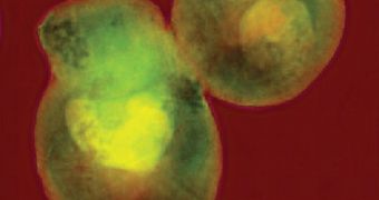
Scientists at the US Department of Energy’s (DOE) Lawrence Berkeley National Laboratory (Berkeley Lab) announce that they were able to use X-ray diffraction microscopy to snap images of whole yeast cells for the first time. The method was applied with an unprecedented resolution, of only 11 to 13 nanometers, and represents a world record for analysis of specimens of this type. The innovation was achieved at the Berkeley Lab Advanced Light Source's (ALS) beamline 9.0.1.
The major implication of the new work is the fact that, fairly soon, researchers could be able to conduct three-dimensional (3D) tomography of single cells at resolutions equivalent to this one. The ALS beamline 2.1, called the National Center for X-Ray Tomography, already performs such measurements, but only at resolutions of 40 to 60 nanometers. The work there is being conducted under the directions of Berkeley Lab Physical Biosciences Division expert Carolyn Larabell. What experts are trying to achieve is the capacity to analyze single cells in their natural, watery environment, and not after dehydration.
“We have demonstrated that lensless imaging techniques can achieve very high resolution while overcoming the limitations of X-ray optics – limitations that include requiring 20 to 50 times the radiation exposure to get a magnified image of the sample. While at present it takes us a long time to image a single specimen – and full 3D imaging of hydrated cells will take even more work – this is a big step in the right direction,” explains the designer of the new imaging method, expert Chris Jacobsen. He is currently based at the DOE Argonne National Laboratory (ANL) and the Northwestern University, and held an appointment at the Stony Brook University.
“Ten-nanometer resolution is easy to achieve with an electron microscope. The problem is that electron microscopy is limited to very thin samples, a few hundred nanometers or less – so you can’t use it to look through a whole cell,” says the co-designer of the imaging method, ALS scientist Janos Kirz. “The challenge of the lensless technique is that essentially the preparation and quality of the sample have to be perfect – and, ideally, completely isolated in the beam,” adds ALS beamline 9.0.1 scientist, Stefano Marchesini. This section of the Advanced Light Source has been built specifically to provide the kind of light needed in X-ray diffraction microscopy.
“True 3D of whole, hydrated, frozen cells at this very high resolution is our next step. One ingredient is to rotate the frozen sample in the beam to get more depth information. Another is to scan the beam across the sample,” Jacobsen concludes.

 14 DAY TRIAL //