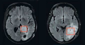
A collaboration of scientists in the United States announce the development of a new imaging technique that allows them to peer deep inside brain cancer cells. This could be of tremendous help in developing therapies against some of the most brutal forms of cancer out there.
Experts focused their attention on gliomas, which are among the deadliest, most common forms of brain cancer. So few treatments and therapies are available against them that the conditions have nearly 100 percent mortality rate.
Investigators with the Massachusetts Institute of Technology (MIT), Harvard University, the Massachusetts General Hospital (MGH) and Agios Pharmaceuticals decided to take matters into their own hands, and concentrate their efforts on the disease’s underlying genetic mutation.
A gene coding for an enzyme called isocitrate dehydrogenase (IDH) has been known to play a role in the development of gliomas for many years, so the team used this as a starting point. About 86 percent of low-grade gliomas display an alteration on this gene.
IDH plays an important role in breaking down sugar molecules inside cells, a process called cellular metabolism. Each cell needs the energy derived from this process in order to survive. Glioma patients can sometimes survive for years.
However, those with high-grade gliomas – referred to as glioblastomas – have poor prognostics. In order to target IDH with drugs, pharmaceutical companies need to have access to an imaging method, which is where the new techniques come in.
The research effort was led by Matthew Vander Heiden, who is a member of the MIT David H. Koch Institute for Integrative Cancer Research. Details of the study appear in the January 11 online issue of the top journal Science Translational Medicine.
According to the research team, applying this magnetic resonance (MR) spectroscopy-based approach to patients could reveal whether their gliomas indeed display IDH gene expression abnormalities.
“The most exciting thing about this is it opens up the possibility that as drugs against gliomas come online, you could know which patients with brain tumors to put in the clinical trials, and you would know if the drug you’re giving them is actually doing what it’s supposed to do,” Vander Heiden says.
The expert holds an appointment as the Howard S. and Linda B. Stern Career Development Professor of Biology at MIT.

 14 DAY TRIAL //