Experts from the Fred Hutchinson Cancer Research Center in Seattle recently managed to gain more insight into how cellular division works, after they analyzed several key components that make the process possible and largely error-free.
There are numerous control mechanisms and checkpoints that cells need to undergo before they can divide into two identical ones. This work flow has been studied extensively, and new advancements in science are allowing experts to go even deeper.
In the new experiments, expert Bungo Akiyoshi, the leader of the research team, managed to study molecules and complex associated with cell division in vitro, demonstrating that they can exhibit the same type of behavior that they do in vivo.
The US National Science Foundation (NSF) provided partial funding for this research, which is detailed in the November 24 issue of the esteemed scientific journal Nature.
“Purifying this molecule out of the cell is as exciting today, as seeing the ribosome was back over 50 years ago!” explains principal study investigator Sue Biggins, who is based at the FHCRC.
One of the main things that scientists known about cell division is that the process is extremely precise, well-synchronized, and thoroughly controlled through a variety of failsafe mechanisms.
The reason why evolution paid this much attention to one event is that cell division can easily turn into an organism's enemy. Failures in this process can either fail to replenish the number of cells in the body, or promote the development of cancer cells and tumors.
In one of the stage of division, genetic material is pooled in the middle of the cell, and then organized into chromosomes. The next step is separating these structures, so as to obtain two complete sets of each.
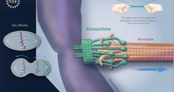
This is done by structures resembling paired microtubules, which radiate from opposite poles of the dividing cell. They perform this action with the aid of a kinetochore, which is a massive molecular complex that is tremendously precise.
The complex can only attach to centromeres, which are spots on the chromosomes that are designed specifically for this purpose. The kinetochore always has a firm grip of the genetic material.
As tension in the microtubules increases, the grip of the molecular complex becomes even tougher. Researchers compare the phenomenon to a Chinese finger trap, which becomes tighter as it's pulled apart.
Experts now believe that the amount of pressure kinetochores use to hold chromosomes may play a significant role in a successful cell division cycle.

 14 DAY TRIAL //