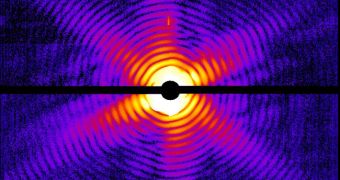
Experts in the field of structural biology are proud to announce a momentous achievement in their area of expertise. Colleagues from an international research group were recently able to use the world's first free electron laser (FEL) to capture an image of an intact virus.
In addition to viewing the tiny microorganism, the FEL was also capable to take a sneak peak at the membrane structure of a photosynthetic species of bacteria. This was done via the use of extremely intensive and ultra-short X-ray pulses.
FEL are innovative, new devices that have never been used in structural biology. The new research effort and its accomplishments are published in two papers that appeared in the February 2 issue of the top scientific journal Nature.
The main implication of the new work, and the reason experts are so happy about it, is that viruses, individual cells, cell organelles, and living bacteria could soon be imaged at the same resolution on a regular basis. Experts never had access to this capability before.
If labs around the world were outfitted with FEL, then studies of biological structures at the molecular level could finally provide answers to some of the most burning questions about microorganisms.
In addition, the group behind the new work argues, the X-ray technique is a lot more efficient than even the most powerful microscopes of today, which means that not even the tiniest biological molecules will escape its keen eye.
“Biologists have long dreamed of being able to capture the image of viruses, single-cell organisms, and bacteria without having to section, freeze, or stain them with metals, as is necessary in electron microscopy,” explains scientist Janos Hajdu.
“Our studies show that it is really possible to create images with the aid of extremely intensive and ultra-short X-ray pulses that would otherwise destroy everything in their path,” he adds.
The study group member was the co-director of the international research team. He holds an appointment as a professor with the Division of Molecular Biophysics at the Uppsala University.
Henry Chapman, also from the university, was the other co-director of the study. Also a part of the research group were experts from the Swedish University of Agricultural Sciences (SLU), led by expert Inger Andersson.
The work is being conducted at the Stanford Linear Accelerator Center's (SLAC) Linac Coherent Light Source (LCLS), the first hard X-ray free electron laser facility in the world.

 14 DAY TRIAL //