At this time, damage inflicted on the heart muscles, or on other tissues surrounding the organ, are very difficult to treat, because of the heart's delicate nature. For years, scientists have dreamed of creating methods of aiding the natural heart-healing process, and experts at the Duke University have recently taken a step forward in that direction – by creating a new type of heart patch. The scientists have also learned how to mimic the developmental process in which embryonic stem cells turn into heart muscle. Their innovation could be used in a large number of heart conditions, healthcare experts say.
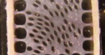
Heart-muscle cells known as cardiomyocytes were the primary target of the new mouse study. In order to create the patch, Duke bioengineers first designed a proprietary 3D mold, on which to grow the cardiomyocytes. The resulting formation acted like the heart muscle should in two distinct and promising ways – it had the ability to contract, and the ability to conduct electrical impulses. This artificial environment, the Duke investigators say, is very similar to the natural conditions of average heart cells.
The research has also determined that helper cells known as cardiac fibroblasts play a huge role in the cardiomyocytes' ability to form lasting heart-muscle cells. The blood-clotting protein fibrin was also added to the 3D mold, to provide mechanical support for the other cells.
“If you tried to grow cardiomyocytes alone, they develop into an unorganized ball of cells. We found that adding cardiac fibroblasts to the growing cardiomyocytes created a nourishing environment that stimulated the cells to grow as if they were in a developing heart,” DU Pratt School of Engineering graduate student Brian Liau says.
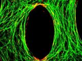
“When we tested the patch, we found that because the cells aligned themselves in the same direction, they were able to contract like native cells. They were also able to carry the electrical signals that make cardiomyocytes function in a coordinated fashion. The addition of fibroblasts in our experiments provided signals that we believe are present in a developing embryo,” Liau adds.
“While we were able to grow heart muscle cells that were able to contract with strength and carry electric impulses quickly, there are many other factors that need to be considered. The use of fibrin as a structural material allowed us to grow thicker, three-dimensional patches, which would be essential for the delivery of therapeutic doses of cells. One of the major challenges then would be establishing a blood vessel supply to sustain the patch,” DU Assistant Professor Nenad Bursac, whose laboratory was used for the new study, shares.
The finds were detailed by Liau at the annual scientific sessions of the Biomedical Engineering Society, held in Pittsburgh. The experiments were made possible through funds provided by the National Institutes of Health (NIH), the National Heart Lung Blood Institute (NHLBI) and the DU Stem Cell Innovation program.

 14 DAY TRIAL //

