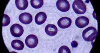
For investigators working to find cures for blood-cell-morphology conditions, such as malaria and sickle-cell disease, gaining a deeper understanding of how the membranes of red blood cells work is of the utmost importance. RBC are among the most peculiar cells in the human body. They do not contain the intracellular structures of other cells, including DNA, but exhibit a high degree of flexibility and resilience. Using a new imaging technique, experts at the University of Illinois now show an advanced method of analyzing the RBC membrane.
Diseases affecting the RBC are generally fatal, for a very simple reason. The donut-shaped cells contain vast amounts of hemoglobin, the molecule that binds to the oxygen we breath into our lungs, and then releases it throughout the body, fueling individual cells. Conditions affecting these blood cells usually imply that hemoglobin can't do its job properly, which in turn means that cells are deprived of oxygen, and that the entire body goes through a slow process of asphyxiation. In order to deliver oxygen everywhere, RBC need to pass through capillaries, the blood vessels connecting veins and arteries, which are sometimes half their own diameter.
Due to their amazingly-constructed membranes, which are both very resistant and highly-deformable, the cells can pass through the capillaries. What the UI team, led by electrical and computer engineering professor Gabriel Popescu, wanted to know was how their ability to perform such “stunts” is related to their morphological traits. “The deformability of red blood cells is their most important property. What we wanted to find is, how does deformability relate to morphology?” explains Popescu, who also holds an appointment at the UI Beckman Institute for Advanced Science and Technology. Details of the study appear in the latest issue of the esteemed journal Proceedings of the National Academy of Sciences.
At this point, biologists have very limited knowledge on the mechanics of the membrane surrounding RBC. The team therefore turned to an innovative imaging technique called diffraction phase microscopy (DPM), in order to gain a better understanding of the factors of play at this level. The thing that separates this method from other types of microscopy is the use of two light beams, rather than the usual one. “One beam goes through the specimen and one beam is used as a reference. It is very, very sensitive to minute displacements in the membrane, down to the nanoscale,” Popescu explains.
In addition to observing nanoscale membrane fluctuations in live cells, the UI team also managed a record first – to measure these fluctuations quantitatively. “An advantage to studying red blood cells in this way is that we can now look at the effects of chemical agents on membranes, specifically. It is very exciting. For instance, we can look at the membrane effects of alcohol, and we may learn something about tolerance to alcohol,” concludes UI College of Medicine instructor Catherine Best, also a coauthor of the PNAS paper.

 14 DAY TRIAL //