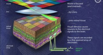
In a groundbreaking new study, researchers have finally been able to map the distribution of signals in the neural circuity that connects individual photoreceptor cells with retinal ganglion cells.
The former type are the ones responsible for carrying electrical signals from the eye to the brain. The retina is the part of the eye that converts incoming light to electrical impulses.
According to the research group, mapping the distribution was only made possible by comparing a clearly defined visual input with the electrical output of the retina.
The accomplishment was made by scientists at the Salk Institute for Biological Studies, whose work was funded by the US National Science Foundation (NSF).
Details of the the work, and of the established neural pathways, appear in the October 7 issue of the esteemed scientific journal Nature.
“Nobody has ever seen the entire input-output transformation performed by complete circuits in the retina at single-cell resolution,” explains expert E.J. Chichilnisky, PhD.
“We think these data will allow us to more deeply understand neuronal computations in the visual system and ultimately may help us construct better retinal implants,” adds the scientist.
Chichilnisky, who holds an appointment as an associate professor in the Systems Neurobiology Laboratories at Salk, was also the senior author of the new paper.
The new document also sheds some light on what type of neural “code” retinal cells use to convert various colors in the light into electrical impulses for the brain.
This investigation was only made possible by the unique neural recording system that the team used. The observational technique was used to record the small electrical signals that numerous retinal neurons generated at the same time.
The methods was developed by high-energy physicists from the University of California in Santa Cruz (UCSC), the AGH University of Science and Technology, in Poland, and the University of Glasgow.
“Just by stimulating input cells and taking a high density recording from output cells, we can identify all individual input and output cells and find out who is connected to whom,” Chichilnisky explains.
Additional funding for the new research came from the US National Institutes of Health (NIH), the Sloan Foundation, and the Engineering and Physical Sciences Research Council (EPSRC), among others.

 14 DAY TRIAL //