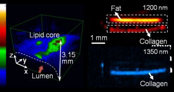
A team of experts at the Purdue University announces the development of a new medical imaging technology, that could be used to diagnose cardiovascular conditions, as well as other diseases.
The method works rather simply, by exposing molecules of interest to very fast-pulsing laser light, and then measuring the ultrasound signals that the molecules produce as a result of their excitation.
One of the most interesting applications for the new technique could be for doctors to gain the ability of analyzing arterial plaques in great detail. These structures can affect blood flow, but soon experts will be able to analyze them in 3D, allowing for more accurate diagnostics and risk assessments.
Purdue associate professor of biomedical engineering and chemistry Ji-Xin Cheng, a member of the research team, says that this approach allows experts to penetrate deep within tissue that would otherwise remain unaccessible.
Other molecular information-gathering methods do not allow for an in-depth analysis, let alone a 3D analysis, but the new one does. As it is adopted around the world, doctors will be able to make more accurate diagnostics, which could in turn help save lives.
“You would have to cut a cross section of an artery to really see the three-dimensional structure of the plaque. Obviously, that can't be used for living patients,” Cheng explains. Details of the new work will appear in the June 17 issue of the esteemed journal Physical Review Letters.
The expert explains that the imaging technique allows the visualization of carbon-hydrogen bonds that keep together the lipid molecules making up arterial plaques. But the method can be tailored to view the fat molecules in muscles to diagnose diabetes, among other things.
“Being able to key on specific chemical bonds is expected to open a completely new direction for the field,” Cheng explains. He and PhD student Han-Wei Wang have been studying this for 4 years.
“We are working to miniaturize the system so that we can build an endoscope to put into blood vessels using a catheter. This would enable us to see the exact nature of plaque formation in the walls of arteries to better quantify and diagnose cardiovascular disease,” Cheng explains.
The 3D images are produced by measuring the time delay produced between the moment the laser light strikes the molecule and the moment when ultrasound waves are detected. This difference can be used to measure distances in a very precise manner, creating a 3D map of the interior of arteries.
“You do one scan and get all the cross sections. Our initial target is cardiovascular disease, but there are other potential applications, including diabetes and neurological conditions,” Cheng concludes.
The new study was funded by grants secured from the US National Institutes of Health (NIIH) and the American Heart Association (AHA).

 14 DAY TRIAL //