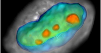
A research group at the Arizona State University (ASU) was recently awarded a $1 million grant for the development of a next-generation, 3D imaging microscope known as a Cell-CT scanner.
The money, provided by the W. M. Keck Foundation, will be used to create a machine capable of performing functional computed tomographic (CT) imaging of individual cells in the living body.
Deirdre Meldrum, an expert with the ASU Biodesign Institute, was awarded the funds, and she will be the leader of the investigations team that will attempt to construct a prototype Cell-CT scanner.
One of the most important feats the new technology will be capable of is analyzing the metabolic pathways of diseases such as cancer as they are used, and not only via biopsies.
This will allow experts to gain new insight into the causes that underlie the development of such dangerous conditions. Studying living cells and assessing their function “live” has been something scientists have been trying to do for many years.
Meldrum’s ASU team will collaborate with scientists at VisionGate, Inc., who will be providing the necessary technology for constructing the scanner. Conceivably, this collaboration will provide healthcare experts with a new tool for medical diagnostics.
“We’re tremendously excited by the potential this technology presents for important breakthroughs, not only in cellular biology but also in medicine and ultimately personalized health care,” says Meldrum.
Experts with the ASU Center for Biosignature Discovery Automation (CBDA) will also be involved in designing the scientific background for the Cell-CT scanner technology.
One of the largest obstacles in creating the device is finding a way of rotating living cells without harming them. CT works by taking a multitude of 2D snapshots of an object, and then combining them together in an integrated view.
The new scanner will be more complex to build because it will need to take photos of structures that are very, very small, and also constantly on the move. The team is expected to complete the device within the next 4 years, as per the terms of the grant.
Ultimately, scientists hope to be able to use the scanner to finally establish the correlations that develop between cell structures and function as diseases such as cancer set in.

 14 DAY TRIAL //