Experts at the University of Maryland in College Park (UM) announce a resounding success in their work with brain-signal recordings. They explain in a new paper accompanying their study that they were able to faithfully reproduce hand motions in three dimensions, using electrical signals recorded from the brain via non-invasive procedures. The group explains that this work could have considerable implications for developing new brain-computer interface systems that are both easier to use and portable. Details of their work appear in the March 3 issue of The Journal of Neuroscience.
“Our results showed that electrical brain activity acquired from the scalp surface carries enough information to reconstruct continuous, unconstrained hand movements,” UM neuroscientist Jose Contreras-Vidal, PhD, the leader of the research team, explains. He says that, until now, many research groups around the world have gone about collecting electrical signals from the brain by placing sensors directly on the cortex. The procedures for that were complex, in the sense that a hole had to be drilled in the skull for scientists to gain access to the brain.
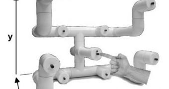
This direct approach does have its advantages. One of them is the fact that the strength of the signal is not muffled by the bones of the skull, as well as by the skin of the scalp. But the UM researchers say that their method produces comparable results, but better functionality. In the new method, an array of sensors is placed on the scalp, and the electrical activity of the brain is recorded via electroencephalography (EEG). In experiments, five volunteers were hooked up to the machine, and then asked to reach for a central button, or four eight other buttons on a device in a random order. The researchers asked them to do this ten times, while they were decoding the electrical impulse map.
Sensors located over the primary sensorimotor cortex (PSC) were the most faithful in allowing for 3D hand motion reconstruction. The PSC is directly involved in controlling voluntary motions, the experts say. Data collected from the inferior parietal lobule also proved to be useful. “It may eventually be possible for people with severe neuromuscular disorders, such as amyotrophic lateral sclerosis (ALS), stroke, or spinal cord injury, to regain control of complex tasks without needing to have electrodes implanted in their brains. The paper enhances the potential value of EEG for laboratory studies and clinical monitoring of brain function,” New York State Department of Health Wadsworth Center expert Jonathan Wolpaw, MD, who has not been part of the research, says.

 14 DAY TRIAL //