The expression “blind as a bat” is deeply rooted in scientific truth. These animals can't see even the smallest amount of light, and fly solely based on echolocation. This is a method in which they emit ultrasounds (noises that are pitched too high for the human ear to detect), and then listen to their reflection to understand what kind of landscape they are navigating. Now, a multi-disciplinary approach has finally determined how bats use this natural ability.
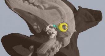
The new work was conducted at the Robarts Research Institute in London, Ontario, by researchers at the University of Western Ontario (Western). They used state-of-the-art micro-computed tomography systems for their investigations, a non-destructive technique that was able to produce 3D scans of the internal anatomy of several types of bats. According to the group, 26 different species of bats, representing about 11 different evolutionary lineages, were analyzed using this method. The main target for tomography was the bone that connected the larynx to the bones that supported the eardrums.
In some bat species, it was determined that the larynx was the preferred method of generating echolocation signals, whereas other bats would rather use clicks generated with their tongues to achieve the same goal, the team says. Details of the research will appear in a paper entitled “A Bony Connection Signals Laryngeal Echolocation in Bats,” which is scheduled for publication in an upcoming issue of the respected scientific journal Nature.
“This discovery may change the way that researchers interpret previous observations from the fossil record of bats. These new results give researchers working with fresh or fossil material an independent anatomical characteristic to distinguish laryngeally-echolocating bats from all other bats,” Western biologist Brock Fenton, the leader of the study, says. The team that made the observations featured imaging scientists, biophysicists, biologists, physiologists, and neuroscientists, from five research institutes all around the world.
“The micro-imaging equipment used in this study was developed in London, Ontario for use in medical research, but it was very exciting to see it used so effectively in basic biological research,” David Holdsworth, who is an imaging scientist at Robarts, adds. “This work is an important step forward in echolocation research because for years, scientists have been searching for a mechanism that would allow echolocating animals to a have a neural representation of their outgoing biosonar sounds for future comparison with reflected echoes of the sounds, and this anatomical discovery may be that mechanism,” McMaster University expert Paul Faure concludes. He provided additional expertise in mammalian physiology and neurobiology for the team.

 14 DAY TRIAL //