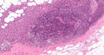
Finding cells in the cornea that may develop into cancerous cells is now possible, announces a collaboration of researchers from Japan and the United Kingdom.
The team says that precancerous cells can be identified by using absorbance spectra data. Having this ability could mean the difference between blindness and sight for thousands around the world.
Cornea tumors respond relatively well to treatment, if they are detected in time. If they are undiscovered, and left untreated, they can become aggressive, extend, and cause loss of vision.
One of the main courses of treatment against this disease is removing the affected cells altogether. But instances in which surgeons remove all cells with the potential to become cancerous are rare.
This makes this a form of cancer that relapses very often, endangering the sight of thousands. By applying the new method, precancerous cells could be identified before surgery is carried out.
Surgeons will from now on be able to detect all dangerous cells, and remove them in one go, therefore reducing patients' risks of relapse, Chemistry World reports.
What the research team did was employ a new approach to scanning the cornea. The team was led by University of Lancaster expert Frank Martin.
The group used synchrotron-based Fourier-transform infrared microspectroscopy as a method of creating an advanced map of the cornea, which can show the different types of cells in the tissue.
The technique can also highlight prospective biomarkers, because it represents a sensitive and high-throughput observations method.
“In the future, this research may well have a direct impact on the diagnosis and treatment of cornea cancer patients,” Martin believes.
“However, more immediately it will have a larger impact in the field of spectroscopy and stem cells due to the potential of the technique for characterization and identification of cancer and disease diagnosis, and in the field of regenerative medicine,” the expert adds.
“Understanding the nature, properties and occurrence of tumor propagating cells, the so called cancer stems cells, is considered vital for the development of new more effective cancer therapies,” says scientist Phil Heraud,
“The work demonstrates synchrotron IR microspectroscopy to be a powerful new phenotypic probe for these studies,” adds the expert, who specializes in cellular biochemistry analysis at the Monash University in Australia.
Details of the new investigation appear in the latest issue of the esteemed scientific journal Analyst, which specializes in publishing high impact research in analytical, bioanalytical and detection science.

 14 DAY TRIAL //