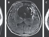
A case report published in yesterday's issue of The New England Journal of Medicine details the removal of what medical experts call a mega-giant aneurysm from the frontal lobe of a 55-year-old man in Boston, Massachusetts.
As explained by specialist Nirav Patel, a neurosurgeon at the Boston Medical Center, aneurysms are labeled giant when they measure more than 2.5 centimeters (1 inch) across.
The one removed from the 55-year-old's brain, however, was much larger than this. Thus, its diameter was one of about 7 centimeters (2.75 inches). In an interview, neurosurgeon Nirav Patel said it was about the size of a plump peach.
The aneurysm triggered personality changes
The 55-year-old man specialist Nirav Patel and his colleagues at the Boston Medical Center operated on was admitted to hospital in January 2013.
The patient sought medical help not because he was experiencing severe headaches or anything of the kind, but because his vision in his right eye had worsened with no obvious cause. Besides, his family had noticed changes in his mood and personality.
Specifically, the man had become forgetful and was having trouble conducting himself in society. Unaware of the aneurysm pressing on his brain, his family simply assumed that he had taken up drinking and that this was causing him to behave erratically.
When the man was examined, however, it was discovered that an artery in his frontal lobe had ballooned and formed a ginormous mass that was pressing on his brain.
“A 55-year-old man presented with a 3-year history of visual impairment associated with personality changes,” reads the medical report on this case.
Furthermore, “Computed tomographic (CT) angiography and magnetic resonance imaging of the brain revealed a partially thrombosed, calcified, 7-centimeter aneurysm of the anterior communicating artery, with surrounding edema.”
The symptoms disappeared following surgery
The massive aneurysm pressing on the 55-year-old's frontal lobe was removed by medical experts in surgery. The intervention lasted around 23 hours, seeing how surgeons not only removed the mass but also repaired the weakened blood vessel.
Shortly after, the man's condition improved to a considerable extent. He got his memory back, albeit with some occasional glitches, and returned to his usual self. He was examined again earlier this year, and doctors found no evidence of the aneurysm making a comeback.
“The patient recovered from surgery and had improvement in his neurocognitive deficits and vision, and he was able to return to work,” neurosurgeon Nirav Patel and colleagues write in The New England Journal of Medicine.

 14 DAY TRIAL //

