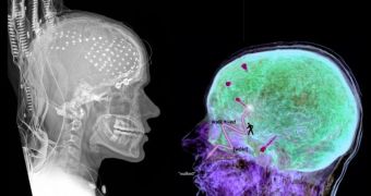
A group of scientists has recently taken a new, high-resolution set of readouts on the human brain, collected directly from the cortex via wires implanted in the brain. The initiative has provided the scientists with an unprecedented look at how language processing functions, and has also evidenced the fact that parts of the processing center are also involved in multi-tasking. The data has been collected from the brains of patients suffering from epilepsy, Wired reports.
“If the same part of the brain does different things at different times, that’s a thunderously complex level of organization,” University of California in San Diego (UCSD) cognitive scientist Ned Sahin says. He has been the leader of the research team that has analyzed a brain region known as Broca’s center, which was known for a long time to be involved in speech and language. The region draws its name from French anatomist Paul Pierre Broca. Details of the investigation appear in the Thursday edition of the top journal Science.
In 1865, Broca was the first to observe that people with damage to this region of the brain lost their ability to speak, but were still able to think and exhibited little other side-effects. Other scientific studies, carried out to test and prove Broca's research, showed pretty much the same thing, but none of them managed to provide a satisfactory explanation of exactly why and how the correlation between speech and this brain area occurred.
Sahin's team did not use the standard functional Magnetic Resonance Imaging (fMRI) viewing technique that other groups did, as it can only hint at which regions of the brain are activated at set time intervals, but not at what goes on inside. Rather, they employed intra-cranial electrophysiology (ICE), a technique that allowed them to position electrodes inside the brain itself, without damaging it.
Participants are “just sitting in a hospital bed, looking at a laptop, and they’re jacked in, with wires right into their brain. And we’re listening to the brain cells talking. It’s fantastic that we cold get so close to the actual neural data. Compared to fMRI, it’s like a close-up, high-speed camera where you can see each beat of a hummingbird’s wings, versus taking a picture of the bird flying around a flower,” Sahin explains.
Patients were made to repeat words daily, and then to place them in past and future tenses. ICE revealed that a small region of Broca's area was responsible for receiving the words, setting them in the requested form, and then sending them back to the speech centers, for articulation. This hints at two uses or more for the brain area. “It’s very distinct from a model where part A does job A. Instead it’s part A doing jobs A, B and C,” the expert adds. “I’m happy to contribute a piece to the puzzle. And the puzzle seems to get more complicated each time you put another piece into it,” he concludes.

 14 DAY TRIAL //