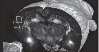
A collaboration of scientists from the Cold Spring Harbor Laboratory (CSHL), in the United States, announces the development of a new imaging technique, which allows investigators to take high-detail anatomical images of the whole mammalian brain.
Thus far, this ability was only the prerogative of very few, highly-specialized research groups that have access to expensive equipment. However, even for these investigators, progress has been slow, since imaging the entire brain is extremely expensive and slow.
What CSHL neuroscientists hope to accomplish is develop a way of circumventing these issues. They want to bring whole-brain imaging to all research groups interested in this topic, so that efforts to fully map the human brain can proceed at a more alert pace.
In collaboration with colleagues at the Massachusetts Institute of Technology (MIT), in Cambridge, the scientists automated and standardized a process that allows a two-photon microscope to divide brain samples into sections.
Once this is done at precise spatial orientations, each of the samples can be imaged sequentially, which opens the way for easier, cheaper and faster whole-brain mapping efforts. The research team was led by CSHL associate professor Pavel Osten.
“The new technology should greatly facilitate the systematic study of neuroanatomy in mouse models of human brain disorders such as schizophrenia and autism,” the team leader explains. He adds that Cambridge, Massachusetts-based TissueVision played an important role in this research.
Details of the imaging technique were published in the January 15 online issue of the top scientific journal Nature Methods. The approach is named Serial Two-Photon Tomography (STP tomography).
“The technology is a practical one that can be used for scanning at various levels of resolution, ranging from 1 to 2 microns to less than a micron,” Osten explains, adding that collecting such a batch of data will now take only 24 hours.
Previous high-resolution, whole-brain imaging techniques required a technician to spend as much as week collecting a similar batch of data, so the new method reduces the amount of time needed for this by 700 percent. This type of innovation is precisely what is needed to move forward, experts not involved in the study comment.

 14 DAY TRIAL //