Viruses may be tiny, but it's not hard to see their effects, when you get sick you notice. Still, actually being able to see them is very useful to researchers.
A couple of researchers at the Australian National University’s School of Molecular Bioscience set about taking a snapshot of a virus as it infects live cells.
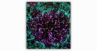
What you see above is the result. It's no simple photograph, it's actually a composite of several shots taken via a microscope.
What's more, the researchers used several techniques to get it looking like it does, not just so it looks cool, but to be able to make out what's happening.
They infected monkey kidney cells with the vaccinia virus, the one used as a vaccine against smallpox, hence the name, and let it be for three days.
Then, they chemically treated their sample and used fluorescent chemicals to make the different parts stand out. What you see in pink are the virus infected cells.
Turquoise is used for the rest of the cells, DNA is yellow. You can notice that the pink cells look different than the healthy ones as most of them are dead or dying, being ravaged by the virus.

 14 DAY TRIAL //