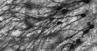
Researchers in the United States announce the discovery of a new signaling pathway in the human brain, that may be responsible for the onset and development of conditions such as mental retardation, epilepsy, and even brain-tumor metastasis.
The team, based at St. Jude Children's Research Hospital, say that they studied key components in the pathway, which allowed them to determine that it controls the movement of neurons through the brain.
Nerve cells generally form in special niches, from where they head out into the cortex, migrating to their final destinations, in the two hemispheres and specialized nuclei. It could be that malfunctions in this pathway could be linked to the development of abnormal brain structures.
The very architecture of the brain is affected when this happens, the research team explains, and defects in this organization are the main cause for many mental disorders and diseases.
In a paper appearing in the November 25 online issue of the esteemed journal Science, the group argues that the finding could be used to make sense of similar mechanisms that are at work in other parts of the body, such as for example in the epithelial tissue.
“Neurons are born in germinal zones in the brain, and the places they occupy in the mature brain are sometimes quite a distance away,” explains team member David Solecki, PhD.
“The cells have to physically move to get to that final destination. If the process is compromised, the result is devastating disruption of brain circuitry that specifically targets children,” he goes on to say.
Solecki holds an appointment as an assistant member of the St. Jude Department of Developmental Neurobiology, and he was also the senior author of the Science paper.
For the new experiments, the team looked at how neural cells departed from their respective germinal zones, and analyzed the molecular complexes that were in competition for controlling this process.
One of the things that allowed for the breakthrough was the development of a fluorescent probe, that was used in combination with an imaging technique called time-lapse microscopy.
This allowed scientists to view the cell-to-cell binding process in real-time. “Until now, cell adhesion was difficult to detect and the techniques involved were laborious,” Solecki explains.
“With this approach, it is almost as if the cells are telling us what they are doing. It was very exciting for me to look at a dish of living neurons and see adhesion occur for the first time,” he adds.
The new work was funded by the National Cancer Institute (NCI), the March of Dimes and ALSAC, AlphaGalileo reports.

 14 DAY TRIAL //