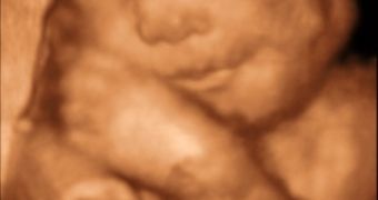
The latest fashion among young parents is to have pictures not only with your new-born baby, but also from before his birth. When the kid grows up, mom will tell him: "Look, this is you when you were...well...uh...minus three months old."
The next big thing will be 3-D movies with the unborn baby, along with the fishing trips, school plays and graduation, to be watched over and over by mom and her senior neighbor friends.
According to researchers at Duke University's Pratt School of Engineering, this could soon be possible, as parents-to-be might soon use 3-D glasses in the ultrasound lab to see their developing fetuses in the womb in living 3-D, just like at the IMAX movies.
After first developing real-time, three-dimensional ultrasound imaging, the same scientists created an updated version of the image-viewing software found on clinical ultrasound scanners, making it possible to achieve a stereo display with no additional hardware, with a modified commercial version of the scanner.
"To our knowledge, this is the first time it's been made possible to display real-time stereo image pairs on a clinical scanner," said Stephen Smith, a professor of biomedical engineering at Duke. "We believe all 3-D scanners could be modified in this way with only minor software changes."
Our depth perception is due to the stereo vision - the slightly different perspectives of the same scene that are observed by the left and right eyes -the way the brain processes the information to produce a sense of depth, a phenomenon that can't be achieved when viewing a single, flat image.
By taking two "snapshots" of the same object from slightly different angles, mimicking the normal difference between left and right eye views, one can create the illusion of three dimensional imaging, just like in the theaters where they give out the funny-looking red and blue glasses.
The scientists, in order to demonstrate the new capability, first generated stereo ultrasound images of a small metal cage. They then advanced to ultrasound images in living animals of a heart valve and blood vessels and needle biopsies of the animals' brains and esophagi.
The researchers have since recorded ultrasound images of a model human fetus that is traditionally used in the testing of fetal ultrasound imaging applications.
"Thousands of 3-D ultrasound systems in clinics could be upgraded with such new software, and stereoscopic goggles could be issued to them as well," Smith said. "Keepsake DVDs of the fetal exam could also be viewed at home in 3-D stereo."

 14 DAY TRIAL //