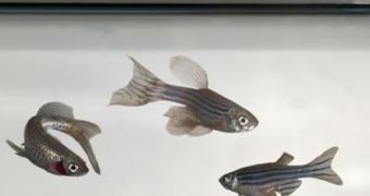
The field of human health research is heavily reliant on the usual monkeys and lab rats, but other animals are used for assessing disease development or drug efficiency as well. Among them are the small fruit fly and the zebra fish. The latter was cloned several times over the last years, but through complex and expensive methods. Now, scientists from the Michigan State University (MSU) have developed a new way of doing this that is both cheaper and more effective than the previous method.
According to a paper published in the latest issue of the journal Nature Methods, for the past 20 years, zebrafish have served as animal models for analyzing human development, as well as defects that may occur at birth. “After the mouse, it is the most commonly used vertebrate in genetic studies. It is used in cancer research and cardiovascular research because they have many of the same genes we have,” MSU Professor of Animal Science Jose Cibelli, one of the co-authors on the new study, says.
Over more recent times, the zebrafish has been used for more complex analyses. For instance, a number of studies has focused on the way certain groups of cells function within the body, and the animal of choice for viewing this was the small fish. With the growing number of uses experts have for it, cloning the animal has become a very important objective, but one that has been achieved only marginally. With the new cloning method, the experts say, the cloning rate success increases to 13 percent, as opposed to the usual 11 percent. This is a very significant difference in the field of bioengineering.
In the new method, an egg is gently removed from a zebrafish and placed in an ovarian solution harvested from a Chinook salmon. “This worked well, because it kept the egg inactive for some time. It gave us two or three hours to work with. It was also useful that we are working in Michigan. The state's Department of Natural Resources was very generous in helping us collect fluid from female Chinooks,” Cibelli, who has been the leader of the MSU team, adds. In the next step, DNA is removed from the egg via a laser, a process adapted from human in-vitro fertilization (IVF).
Then, researchers had to figure out a way of implanting donor DNA in the cell. “The tricky part was finding a way to get into the egg. We used the same entrance that [the male reproductive cell] uses. There was only one spot on the egg, and we had to find it,” Cibelli explains. “So far the mouse is the only one from which you can delete genes in a reliable fashion. what researchers do is mutate a gene, abolish its function completely, and then study the consequences,” the expert concludes, quoted by ScienceDaily.

 14 DAY TRIAL //