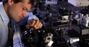
A collaboration of investigators from the United States announces a major breakthrough in imaging technologies used for a variety of medical applications. The group learned how to use optical-coherence tomography (OCT) to image the heart of fetuses inside their mothers' wombs, even before the small organs begin to beat. The technique is non-invasive and produces no ill side-effects, they say.
With the help of the new method, the investigators can now obtain images of life as it begins to take place. This is bound to lead to significant advancements in studying fetuses, as well as progress in doctors' abilities of identifying potential heart problems in the small ones very early on. This is important for their general chances of surviving, as early interventions are often the only way to save their lives.
The new method was developed by University of Houston Cullen College of Engineering assistant professor of biomedical engineering Kirill Larin, who worked together with scientists from the Texas Medical Center. The high-resolution type of OCT they are using allows for them to track the development of mammalian hearts. Therefore, they benefit from the most advanced imaging method of vital organ development ever produced.
“Everything we know about early development of the heart and formation of the vasculature system comes from in vitro studies of fixed tissue samples or studies of amphibian and fish embryos. With this technology, we are able to image life as it happens, see the heart beat in a mammal for the very first time,” the expert explains. OCT is a technology that is heavily based on analyzing the reflection of an infrared laser beam hitting a target object or tissue. The new investigation was conducted with a $1.7 million grant from the US National Institutes of Health (NIH).
"We are using OCT to image mouse and rat embryos, looking at video taken about seven days after conception, out of a 20-day typical mammalian pregnancy. This way, we are able to capture video of the embryonic heart before it begins beating, and a day later we can see the heart beginning to form in the shape of a tube and see whether or not the chambers are contracting. Then, we begin to see blood distribution and the heart rate,” Larin says.

 14 DAY TRIAL //