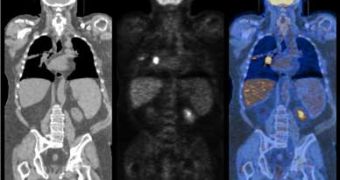
Medicine researchers and physicists are working together to create new electronic components that would enable them to identify subatomic particles in high-energy accelerators with increased accuracy.
Different applications using the same technology could help measure the velocity of subatomic particles much better than before and even detect cancer at an earlier, more curable stage.
"The electronics needs in medical imaging look very closely related to the needs we have in high-energy physics," said Henry Frisch, Professor in Physics at the University of Chicago. "Physics tends to advance by new capabilities in measurement, the same in radiology."
Researchers received initial funding from the U.S. Department of Energy, Argonne and the University of Chicago Cancer Research Center, are trying to apply high-energy physics to biomedical imaging techniques.
The physicists' interest in this technique regards the types of subatomic particles that could be observed in large colliders, since many such particles have only been theorized, but their appearance is still unknown.
The highest accuracy in measuring the speed of these particles is of 100 picoseconds (a trillionth of a second), but considering that a single photon travels about one inch in this time interval, higher resolutions are sought.
With a resolution close to 30 picoseconds, the quality of the image would greatly improve, resulting in more accurate pictures of many subatomic particles, but in order to do this, scientists have to rely on the newest technique, called "time-of-flight PET, which provides a positional measurement that conventional PET technology lacks.
"If time-of-flight measurements can be assessed with an accuracy less than 30 picoseconds, better resolution in both directions can be achieved, essentially eliminating the need for complex and costly image reconstruction," Chen said.
A new microchip may increase the capability to measure the particle velocity. It's 850 microns across, around the same as a dozen human hairs lying side by side.

 14 DAY TRIAL //
