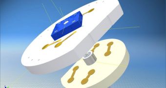
German researchers at the Fraunhofer Institute for Photonic Microsystems IPMS, in Dresden, announce the development of a new tool that could be used to detect cancer in vivo. Generally, when doctors check for tumors or other signs of the disease, they collect a piece of tissue from an area of interest (the process is called biopsy), and then send it to laboratories for analysis. The method is fairly accurate, but it takes a lot of time to perform, and the IPMS team wanted to develop a technique of detecting cancer live, as doctors were investigating their patients.
In their experiments, the scientists came up with an innovative approach of doing that. They developed an advanced type of microscope head, which they say can make out, and then magnify, tissue samples just 10 to 20 micrometers in diameter. Additionally, the head itself is just 8 millimeters in diameter, which means it can easily be used inside the human body, most likely attached to an endoscope. Healthcare experts could, therefore, spend a lot more time looking around, rather than being confined to harvesting only a limited amount of samples. With the new approach, detecting cancer could become a lot faster, and also more precise.
Detecting the disease in its earliest stage is crucially important, if the patient is to have any chances of surviving their ordeal. If doctors discover cancer when the condition has already metastasized, then it's generally too late for that individual. On the other hand, not ever lump that appears on the human body is a malignant tumor, so what researchers really needed was a method of conducting extensive surveys of such structures, without putting the patients through the discomfort of biopsies. The time samples usually spend in scientific labs is also eliminated, which means that tumors, if they exist, are found, and then addressed, a lot faster than they would have been otherwise.
“Microscopic image recorders that can be used on endoscopes have not been available up to now. We have developed the first laser-based sensor for this purpose. In classic endoscopy using macroscopic imaging, the job can be done by CCD or CMOS image sensors, as used in digital cameras and cellphones. For endomicroscopy, however, MEMS-based image sensors are highly advantageous because they can magnify even the smallest object fields, such as cells, without the need for a large lens. We have combined the sensor with a microscanner mirror to achieve the required resolution of 10 micrometers and can therefore massively magnify the tiniest structures,” explains IPMS business unit manager Dr Michael Scholles.
“An important aspect of the development was to produce a suitable microassembly for the endoscope head. Here we faced the challenge of making the complete system suitable for installation in the endoscope, and we managed to do it. In future our microscope head will be produced in large quantities in an automated process for subsequent installation in endoscopes. [The system] could be used not only in medical and biological microscopy but also in technical endoscopy, for instance to examine cavities in buildings or to inspect the insides of engines and turbines,” he concludes, quoted by PhysOrg.

 14 DAY TRIAL //