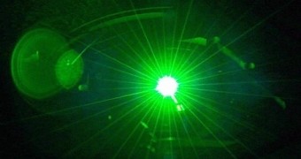
Photo and video cameras used by normal people won't be seeing any huge quality benefits due to the new breakthrough from Yale, but the technology has huge implications for biomedical imaging, microscopes, photolithography and even holography.
Researchers from Yale, a private Ivy League research university in New Haven, Connecticut, have developed a semiconductor laser that merges the benefits of low image corruption inherent in LEDs (light emitting diodes) with the brightness of traditional lasers.
Chaotic cavity lasers didn't actually have many, if any, practical applications until now, just like random cavity lasers (no, they're not the same thing apparently).
Now, it has been established that they can be used to solve some of the largest problems in imaging and microscopy.
Quite a leap really, from a basic sort of research to the golden solution for one of the more troublesome and stifling limitations of man's technological level.
How the new semiconductor laser works
Light sources are as important for microscopy, especially electron microscopes, as the lens arrays used to focalize that light into an image viewable on a screen or through an objective.
One of the biggest issues is speckle, a random, grainy pattern caused by high special coherence. It can corrupt the formation of images when normal lasers are used.
Using LEDs as light sources solves some of the problem, but they are simply not bright enough for high-speed imaging.
That's where the new electrically pumped semiconductor laser comes in. It has a low spatial coherence but nonetheless produces a very intense beam of light.
This leaves the speckle contrast at 3%, which is even better than the maximum 4% needed to avoid any disturbance in the view that eventually reaches our eyes.
Compared to standard edge-emitting lasers which produce a speckle contrast of around 50%, that's a massive improvement. No more coherent artifacts to worry about from now on.
Practical applications
Studying of how tissues and materials work on a microscopic level is a no-brainer, as is biomedical engineering, perhaps, but there are many other, more “mundane” scenarios like pediatrics, radiology and diagnostics.
Certain classes of clinical diagnostics that use light have been out of the question until now, but the avenues of research are open once more.
The breakthrough was detailed in a paper published on January 19 in the online edition of the Proceedings of the National Academy of Sciences.

 14 DAY TRIAL //