Researchers at the Georgia Institute of Technology's (Georgia Tech) George W. Woodruff School of Mechanical Engineering announce the development of a new medical device that is capable of producing 3D maps of the human heart from within the organ itself.
The team is directly responsible for creating the underlying technology for the new catheter-based device. Once inside the human body, the tiny instrument is able to produce real-time, forward-looking images of the heart, potentially helping doctors with both diagnostics and treatments.
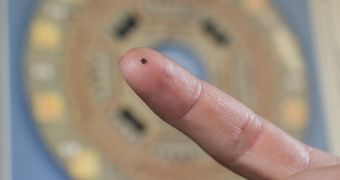
The 1.4-millimeter chip is also able to work from within coronary arteries and peripheral blood vessels, where larger catheters simply cannot be used due to their size. One possible application the device enables is the use of non-invasive procedures to clear arteries clogged by cholesterol.
One of the most interesting aspects of the innovative instrument is that it manages to integrate processing electronics and ultrasound transducers in such a small structure. Furthermore, all processing is conducted on-chip, without the need to relay the data outside of the patients' bodies.
All hundred elements on the chip can therefore synthesize readings in a manner that allows researchers to recover data using as few as 13 cables. Overall, the chip displays better performances when it comes to creating relevant images than any existing cross-sectional ultrasound techniques.
The team, led by Georgia Tech professor F. Levent Degertekin, says that the earliest version of the device was able to capture readings 60 times per second, and adds that better performances will most likely be enabled in future generations of the instrument.
“Our device will allow doctors to see the whole volume that is in front of them within a blood vessel. This will give cardiologists the equivalent of a flashlight so they can see blockages ahead of them in occluded arteries,” he says.
“It has the potential for reducing the amount of surgery that must be done to clear these vessels,” the investigator adds. Additional details of the instrument were published in the February 2014 issue of the journal IEEE Transactions on Ultrasonics, Ferroelectrics and Frequency Control.
The work was supported by the National Institute of Biomedical Imaging and Bioengineering (NIBIB) at the US National Institutes of Health (NIH).

 14 DAY TRIAL //