A group of investigators at the Massachusetts Institute of Technology, in Cambridge, announces the development of a microfluidic device that is able to separate the basic components of a liquid mixture.
The team behind the work says that the tool is very well suited to conducting analyses of human blood, since it can separate the various components from each other quite easily. This means that the device will make for an excellent diagnostics tool in the future.
But human blood is not the only complex chemical mixture that could be analyzed via the new instrument. A wide range of applications awaits it in chemistry, biology and product manufacturing.
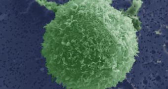
However, it's quite clear that its main use will be in medicine. The microfluidic device is perfectly capable of identifying cancer cells, for example, in a blood sample. In fact, it might be able to do so more precisely and more efficiently than any other method.
Tumor cells, stem cells or fetal cells could easily be identified or separated from regular cells, for whatever applications investigators might devise for them. At this point, doing so is a very complex and energy-intensive process.
“You’re basically looking for a needle in a haystack,” study researcher Sukant Mittal explains. He is a graduate student with the Harvard-MIT Division of Health Sciences and Technology (HST). The MIT group worked closely with colleagues from the Massachusetts General Hospital (MGH) on the study.
Details of the investigation and of the new microfluidic device were published in the February 21 issue of the esteemed Biophysical Journal. The medical instrument utilizes small channels to bring whatever mixtures are being analyzed in contact with antibodies located within.
This approach – using antibodies – is not new in itself. What is new is the method used to direct the flow of liquids towards areas where the target cells the device is looking for can come in contact with their antibodies. The latter then indicate detection.
In previous attempts to use antibodies, the efforts were stifled by the fact that target cells were moving very fast, making it very difficult for the detecting molecules to latch on. Now, the channels in the microfluidic device handle this issue by allowing the test liquid to flow only in specific patterns.
“Considerable validation and testing will be necessary before this early-stage device can be deployed in the clinic. Nevertheless, this novel approach may enable exciting diagnostic and therapeutic opportunities that are not feasible using existing technologies,” study researcher Mehmet Toner adds.

 14 DAY TRIAL //