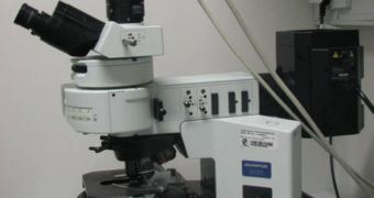
The fluorescence microscope (FM) has been the instrument of a whole generation of scientists over the last years, allowing the researchers to peer at things as small as proteins, moving live inside their containing cells. But, as technology evolved, humans started constructing various structures on the nanoscale, which means that the FM lagged somewhat behind in development. Recently, an engineer at the University of Georgia (UGA) has announced that he has developed the basic principles of a new form of microscopy, which has twice the resolution of FM and makes use of the physical properties of structured illumination.
The paper detailing the finds, published in the April 26th issue of the scientific journal Nature Methods, shows that, while fluorescence microscopy can only image fluorescent markers in the 200-nanometer scale, the new observation method can easily resolve details twice as small, in the 100-nanometer range. With this advancement, experts studying in the field of biosciences believe that they may finally be able to start seeing processes that are going on at a too small a scale to be picked up with existing microscopes. They also hope to get a better glimpse of how diseases affect the cell, and how the latter, as a whole, behaves in certain circumstances.
“Our understanding of what is going on inside cells and our ability to manipulate them has advanced so much that it has become more and more important to see them at a better resolution,” UGA Engineer Peter Kner, who has been the main developer of the new microscopy technique, explained. For the innovation, he collaborated with colleagues from the University of California in San Francisco (UCSF). The experts worked on the basic principles of the fluorescence microscope, which boasts a history of more than 50 years.
“What we've done is develop a much faster system that allows you to look at live cells expressing GFP, which is a very powerful tool for labeling inside the cell,” Kner added. “It would be difficult to overstate the importance of bio-imaging to ongoing research in human health. The ability to shine a light on the leading-edge of scientific discovery will define the route to entirely new regimens of health management at the intersections of science and engineering, and Dr. Kner has joined a distinguished cadre at UGA to continue working at that interface,” the UGA Faculty of Engineering Director, Dale Threadgill, concluded.

 14 DAY TRIAL //