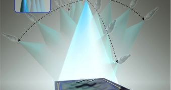
Scientists in the United States announce the creation of a new type of microscope that can be used to create three-dimensional tomographic images of micro- and nanoscale samples. The entire system is at the same time small enough to fit into the user's palm.
But the real kicker is that the instrument doesn't need any sort of lens to carry out its investigations, say researchers from the University of California in Los Angeles (UCLA), where the device was created.
According to the group, this is the first time when the technique known as lens-free optical tomographic imaging is shown to be capable to producing 3D images of multiple microscopic objects, while at the same time providing high resolution capabilities.
UCLA specialists also say that the entire instrument can neatly fit into an opto-electronic chip, which means that it should be able to take advantage of the automation implies by such systems. The immediate benefit is that scientists could complete the same imaging tasks way faster than today.
“This research clearly shows the potential of lens-free computational microscopy,” explains the senior author of the new study, UCLA Henry Samueli School of Engineering and Applied Science associate professor of electrical engineering Aydogan Ozcan.
“Wonderful progress has been made in recent years to miniaturize life-sciences tools with microfluidic and lab-on-a-chip technologies, but until now optical microscopy has not kept pace with the miniaturization trend,” the expert goes on to say.
Cell and developmental biology are expected to be the research fields that will stand most to gain once this technology is employed at a larger scale, at research labs around the world.
An added benefit to constructing microscopes so small, the UCLA experts say, is that important savings are also made in equipment and material costs. Larger, more complex imaging instruments cost tens of thousands of dollars also because they include numerous large components.
“Lens-free imaging removes the pipes altogether by utilizing an entirely new design,” Ozcan explains. The fact that the UCLA team managed this innovation is a sign that now, 400 years after the invention of the microscope, improvements can still be made to this basic scientific tool.
“The field of view of lens-based microscopes is limited because the lens focuses on a narrow area of a sample. A lens-free microscope has both a much larger field of view and depth of field because the imaging is done by the digital sensor array and is not constrained by a lens,” says Waheb Bishara PhD.
The expert is based at the California Institute of Technology (Caltech), in Pasadena, California.
Funds for the study were secured from the US National Science Foundation, the Office of Naval Research and the National Institutes of Health. The Gates Foundation and the Vodafone Americas Foundation also contributed to the research.

 14 DAY TRIAL //