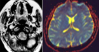
Investigators at the Cambridge-based Massachusetts Institute of Technology (MIT) announce the development of an algorithm that is capable of analyzing the vast volumes of raw data produced by various medical brain imaging techniques, and then combining them into neural maps.
Basically, what the algorithm does is map out the areas that have already been affected by conditions such as schizophrenia, and then figure out how the damage spreads to other portions of the brain.
Having access to this type of information could help doctors better tailor the treatments they propose to patients. In addition, pharmaceutical companies may use the data to design better drugs, capable of causing effects exclusively in areas where they are needed, while leaving healthy nerve cells alone.
Scientists have been trying to gain access to this capability for quite some time now, but piecing together the massive volume of data that medical imaging techniques produces turned out to be impossible, until now.
The MIT team was able to create an algorithm that can piece together information provided by multiple imaging methods, and provide an in-depth view of a patient's brain, and their overall health levels.
Details of how the asset works will be presented at the International Conference on Medical Image Computing and Computer Assisted Intervention, to be held in Nice, France, in October. The team behind the work is based at the MIT Computer Science and Artificial Intelligence Laboratory.
The algorithm proper was developed by MIT associate professor of computer science Polina Golland, and graduate student Archana Venkataraman, and is able to combine data from diffusion and functional magnetic resonance imaging (MRI) datasets.
While dMRI analyzes water diffusion patterns in white matter, fMRI reveals blood flow patterns to various regions of the brain. This is useful because it shows doctors which areas are activated at any given time, or in response to a certain stimuli.
“It’s quite hard for a person looking at all of that data to integrate it into a model of what is going on, because we’re not good at processing lots of numbers,” Golland says. The new algorithm, however, is not subject to such constraints.
“It is based on the assumption that with any disease you get a small subset of regions that are affected, which then affect their neighbors through this connectivity change. So our methods extract from the data this set of regions that can explain the disruption of connectivity that we see,” she concludes.

 14 DAY TRIAL //