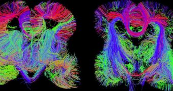
A team of investigators including scientists from the Allen Institute for Brain Science in Seattle announces the development of a new high-resolution map of the human brain, which reveals the most important neural connections long before a fetus is born. This type of in-utero map is unique, and is already being used to investigate the origins of conditions such as autism and schizophrenia.
Some of the most important things revealed on the new map are the locations where particular genes are being switched on or off. Multiple mental disorders are caused by faulty gene expression, or include a genetic component, so having the ability to observe the neural development process as it happens may give scientists a deeper insight into what is causing those issues.
According to the AIBS group, the map was created at the midpoint of pregnancy, a critical time when the fetus is developing essential structures inside their brains. Details of the new study were published in the April 2 issue of the top scientific journal Nature, NPR reports. Having access to in-utero brain maps is important because this organ develops from a single cell into 80 billion cells in just 9 months.
“It's a pretty big leap. Basically, there was no information of this sort prior to this project,” explains AIBS investigator Ed Lein, who was actively involved with creating the new dataset. Even though many psychiatric or behavioral problems do not start manifesting until a person reaches their 20s, the origins of these issues can be traced back to the fetus stage.
The new investigation was conducted as part of the BrainSpan Atlas of the Developing Human Brain, and benefited from federal support in the United States. The work also included scientists with the US National Institute of Mental Health, which is part of the US National Institutes of Health (NIH)
“We're talking about a remarkable process,” explains NIMH director, Dr. Thomas Insel. He explains that the new study involved the team using a laser to cut tens of thousands of brain tissue samples into very tiny slices. All material was collected from aborted fetuses. For each of these tiny slices, experts checked to see which genes were turned on or off.
Lein explains that this was needed in order to determine which types of cells were present in specific regions of the brain. Additionally, the data was used to determine the role that those cells were playing. Already, the new map has been used to make two significant findings, the expert says.
“The first is that many genes that are associated with brain disorders are turned on early in development, which suggests that these disorders may have their origin from these very early time points,” Lein explains. The second discovery is that the human brain is different from the mouse brain in several ways researchers were unaware of until now.
Though it may appear self-obvious, this discovery is actually very important, since numerous drugs are tested on mice that then end up in pharmacies and hospitals around the world. Figuring out when we can use mice as human models, and when we cannot, is extremely important.

 14 DAY TRIAL //