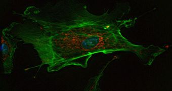
Learning how cells respond to various stimuli is one of the areas of research that have the potential to reveal a wealth of data about biological systems. These pieces of information could inform experts in designing better drugs and therapies for a variety of medical conditions, but a more in-depth study of complex biological systems is still very difficult to perform. Now, a team of scientists from the United States has managed to create a new technique for using images of single cells to identify genetic interactions within biological networks. Experts say that this may be the first steppingstone to the future of high-throughput cell imaging analysis.
This is a vast improvement from the methods applied just a few years ago. For many decades, researchers have been confined to investigating cells through the microscope, and observing the way they react to certain stimuli, but collecting data in this manner is done qualitatively by eye. Recent advancements in technology, however, promoted the development of various high-throughput imaging methods that produced extensive datasets of hundreds of different morphological features of the same cell, in roughly the same amount of time.
Now, based on this ability, researchers at the Massachusetts Institute of Technology (MIT), and the Harvard Medical School, have developed the new method. The goal is to get more details of cell morphology, and the way this is accomplished is by exploring the cellular networks that regulate them. “These images are an enormous source of data that is only beginning to be tapped. We realized we had enough data to go beyond classification and start to understand the mechanism behind the differences in shape,” Bonnie Berger, an MIT scientist, explains. The investigator is also the senior author of a new paper detailing the findings, which appears in the latest issue of the journal Genome Research.
“This work provides a glimpse into the future, where looking under the microscope manually at cells one-by-one is replaced with automated high-throughput processing of many cellular images,” Berger adds. The method could be combined with other means of gathering data, so that the researchers working in this field can benefit from a complete set of tools to collect all the information they need to appreciably increase the accuracy of known signaling networks.

 14 DAY TRIAL //