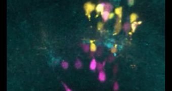
In an investigation conducted on zebrafish, researchers at the University of Cambridge studied the patterns in which the retina develops, gaining new insights into the true complexity of the nervous system. The entire retina formation process was imaged in four dimensions.
This tremendous achievement was made possible by using a series of powerful genetic labeling techniques and advanced neural imaging capabilities. The research group was led by Cambridge professor Bill Harris.
The central nervous system (CNS) comprises not just the brain, but also the spinal cord and the retina. This organ, the most complex in the body, is made up of a vast number of different cell types, all of which are produced from a lump of undifferentiated cells.
Harris, who holds an appointment as the head of the Cambridge Department of Physiology, Development and Neuroscience, and is also an experimental biologist, says he has been fascinated by how this transition occurs for many years.
The expert and his team are the first ever to observe the entire process of retinal development at the cellular level in zebrafish embryos. Their achievement also marks the first time a vertebrate CNS structure was observed as it developed in vivo.
“What surprises me most is the inscrutable logic of it all. Every experiment designed to differentiate between different hypotheses leads down one or other branch of an untraveled winding way, through a complex and cleverly evolved network that eventually leads to the functioning CNS,” Harris says.
“We use the retina as a model for the rest of the brain, and zebrafish are a useful research model species because their transparent embryos are easy to see into, and develop rapidly,” he adds.
While humans take around one year (pregnancy plus a few months after birth) to fully develop their retinas, zebrafish embryos take only three and a half days to do the same. By using a sophisticated confocal microscope, the team is able to observe the process in detail.
“Forty years ago you would look at the embryo and label some cells, then later look again to see what cells were now labeled or where they were,” Harris says.
“You’d get snapshots under the microscope and surmise what happened in between. Now we are actually seeing how the cells divide, which gives us a lot more information to help distinguish certain hypotheses from others,” the expert concludes.

 14 DAY TRIAL //