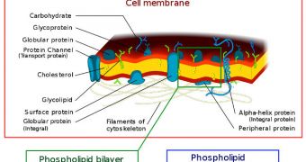
A collaboration of American and Japanese investigators was finally able to shed light on a decades-old mystery when they created movies of single molecules traveling through living cell membranes.
This is the first time this has been achieved in a living system, and now scientists can finally gain a better understanding of how receptor molecules on the membrane behave under certain conditions.
Until now, this was unknown, as methods of imaging the interactions that occur in the membrane of a living cells are something very new. Working together, colleagues from the Kyoto University and the University of New Mexico (UNM) managed to achieve their common goal.
Thanks to their unrelenting work and innovation, scientists now have access to new possibilities for drug development and research, that would have been impossible to attain otherwise.
Details of how the new movies were made were published in the latest issue of the esteemed scientific Journal of Cell Biology, Science Blog reports. Each of the video clips is focused on a single molecule.
The team investigated closely a class of molecules that makes up the largest superfamily in the entire human genome. They are called G protein-coupled receptors (GPCR), and they are the target of nearly half of all modern drug development efforts.
In spite of all this, no one knew until now how these molecules played an important role in relaying signals from the extracellular environment to cellular components past the membrane.
They were known to relay signals, but the exact mechanisms they employed to do so remained a mystery until now. As such, the pathways these molecules use to take care of business have been a subject of debate in the international scientific community for up to 15 years.
What researchers wanted to know was whether these molecules prefer to work on their own, as monomers, or whether they are team players called dimers. The new investigation proved that the truth is somewhere in the middle, confirming and denying both hypotheses at the same time.
“By developing a super-quantitation single-molecule imaging method, in which GPCR molecules are inspected one by one in living cell membranes, we are now able to actually ‘see’ that each individual FPR molecule moves around in the cell membrane, endlessly interconverting between monomers and dimers with different partners, completing each cycle within a quarter of a second,” says Rinshi Kasai.
The expert, who is based at the Kyoto University Institute for Integrated Cell-Material Sciences (iCeMS), is the lead author of the new paper.
“We obtained a parameter called the dissociation constant, which will allow us to predict numbers of monomers and dimers if the total number of GPCRs in a cell is known,” adds expert Akihiro Kusumi.
“The ability of scientists to obtain such key numbers will be essential for understanding GPCR signaling, as well as defects leading to diseases from the neuronal to the immune systems,” he adds.
“The implications for drug design, blocking signal amplification by monomer-dimer interconversion, are profoundly important,” concludes the expert, who is also an iCeMS professor.

 14 DAY TRIAL //